Esoteric Diagnostic Considerations for Small Round Cell Tumors in Biopsy Specimens With Extensive Negative Immunohistochemical Profiles: Utilizing Subtle Histopathological Features Prior to Molecular Testing.
{"title":"Esoteric Diagnostic Considerations for Small Round Cell Tumors in Biopsy Specimens With Extensive Negative Immunohistochemical Profiles: Utilizing Subtle Histopathological Features Prior to Molecular Testing.","authors":"Dong Ren, Ryan O'connell","doi":"10.1155/crom/5186729","DOIUrl":null,"url":null,"abstract":"<p><p>Small round cell tumors (SRCTs) are characterized by primitive round cells and a broad differential diagnosis due to their undifferentiated nature, making their diagnosis particularly challenging. Molecular testing is often essential for definitive classification; however, subtle histomorphological features can significantly narrow the differential diagnosis. Here, we present the case of a 44-year-old male who presented with a painless mass (up to 15.6 cm) in the left thigh. Histologic examination of the biopsy revealed solid sheets of monotonous small round cells with scant cytoplasm, hyperchromatic nuclei, and conspicuous nucleoli within the edematous to myxoid stroma. Notably, capillary-sized blood vessels were present throughout the tumor, which made BCOR-rearranged sarcomas, myxoid liposarcoma with small cell morphology, and GLI1-altered soft tissue tumors the main differential diagnoses. Classic morphology of myxoid liposarcoma was not present. Immunohistochemical (IHC) staining revealed that the tumor cells were diffusely positive for SOX11 but negative for SATB2, CD56, S100, and TLE1. This immunophenotype, combined with the histological findings, strongly suggested a diagnosis of myxoid liposarcoma with high-grade features. Fluorescence in situ hybridization (FISH) analysis confirmed a DDIT3 rearrangement, supporting this diagnosis. We hope this case will enhance pathologists' understanding and recognition of the importance of utilizing subtle histologic features to establish the differential diagnosis and accurately diagnose SRCTs in biopsy specimens prior to molecular testing.</p>","PeriodicalId":9636,"journal":{"name":"Case Reports in Oncological Medicine","volume":"2025 ","pages":"5186729"},"PeriodicalIF":0.6000,"publicationDate":"2025-08-04","publicationTypes":"Journal Article","fieldsOfStudy":null,"isOpenAccess":false,"openAccessPdf":"https://www.ncbi.nlm.nih.gov/pmc/articles/PMC12339143/pdf/","citationCount":"0","resultStr":null,"platform":"Semanticscholar","paperid":null,"PeriodicalName":"Case Reports in Oncological Medicine","FirstCategoryId":"1085","ListUrlMain":"https://doi.org/10.1155/crom/5186729","RegionNum":0,"RegionCategory":null,"ArticlePicture":[],"TitleCN":null,"AbstractTextCN":null,"PMCID":null,"EPubDate":"2025/1/1 0:00:00","PubModel":"eCollection","JCR":"Q4","JCRName":"ONCOLOGY","Score":null,"Total":0}
引用次数: 0
Abstract
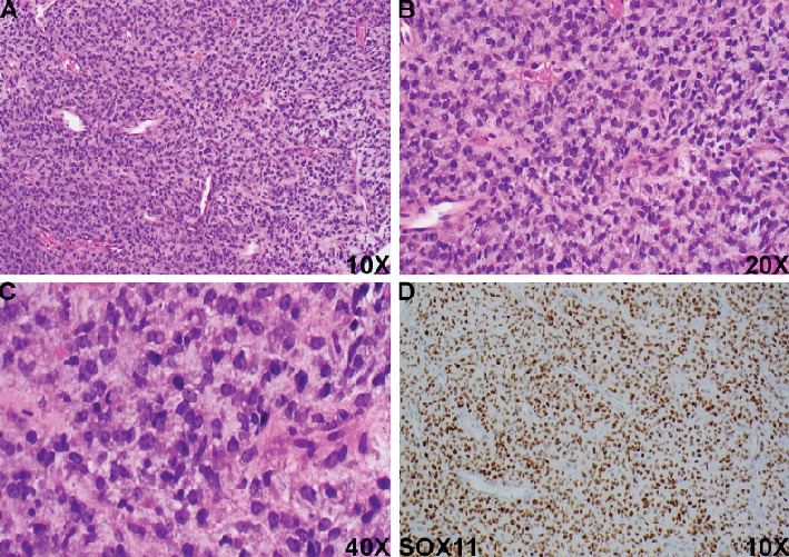
Small round cell tumors (SRCTs) are characterized by primitive round cells and a broad differential diagnosis due to their undifferentiated nature, making their diagnosis particularly challenging. Molecular testing is often essential for definitive classification; however, subtle histomorphological features can significantly narrow the differential diagnosis. Here, we present the case of a 44-year-old male who presented with a painless mass (up to 15.6 cm) in the left thigh. Histologic examination of the biopsy revealed solid sheets of monotonous small round cells with scant cytoplasm, hyperchromatic nuclei, and conspicuous nucleoli within the edematous to myxoid stroma. Notably, capillary-sized blood vessels were present throughout the tumor, which made BCOR-rearranged sarcomas, myxoid liposarcoma with small cell morphology, and GLI1-altered soft tissue tumors the main differential diagnoses. Classic morphology of myxoid liposarcoma was not present. Immunohistochemical (IHC) staining revealed that the tumor cells were diffusely positive for SOX11 but negative for SATB2, CD56, S100, and TLE1. This immunophenotype, combined with the histological findings, strongly suggested a diagnosis of myxoid liposarcoma with high-grade features. Fluorescence in situ hybridization (FISH) analysis confirmed a DDIT3 rearrangement, supporting this diagnosis. We hope this case will enhance pathologists' understanding and recognition of the importance of utilizing subtle histologic features to establish the differential diagnosis and accurately diagnose SRCTs in biopsy specimens prior to molecular testing.
期刊介绍:
Case Reports in Oncological Medicine is a peer-reviewed, Open Access journal that publishes case reports and case series related to breast cancer, lung cancer, gastrointestinal cancer, skin cancer, head and neck cancer, paediatric oncology, neurooncology as well as genitourinary cancer.

 求助内容:
求助内容: 应助结果提醒方式:


