{"title":"Gastrointestinal bleeding detection on digital subtraction angiography using convolutional neural networks with and without temporal information.","authors":"Derek Smetanick, Sailendra Naidu, Alex Wallace, M-Grace Knuttinen, Indravadan Patel, Sadeer Alzubaidi","doi":"10.4274/dir.2025.253134","DOIUrl":null,"url":null,"abstract":"<p><strong>Purpose: </strong>Digital subtraction angiography (DSA) offers a real-time approach to locating lower gastrointestinal (GI) bleeding. However, many sources of bleeding are not easily visible on angiograms. This investigation aims to develop a machine learning tool that can locate GI bleeding on DSA prior to transarterial embolization.</p><p><strong>Methods: </strong>All mesenteric artery angiograms and arterial embolization DSA images obtained in the interventional radiology department between January 1, 2007, and December 31, 2021, were analyzed. These images were acquired using fluoroscopy imaging systems (Siemens Healthineers, USA). Thirty-nine unique series of bleeding images were augmented to train two-dimensional (2D) and three-dimensional (3D) residual neural networks (ResUNet++) for image segmentation. The 2D ResUNet++ network was trained on 3,548 images and tested on 394 images, whereas the 3D ResUNet++ network was trained on 316 3D objects and tested on 35 objects. For each case, both manually cropped images focused on the GI bleed and uncropped images were evaluated, with a superimposition post-processing (SIPP) technique applied to both image types.</p><p><strong>Results: </strong>Based on both quantitative and qualitative analyses, the 2D ResUNet++ network significantly outperformed the 3D ResUNet++ model. In the qualitative evaluation, the 2D ResUNet++ model achieved the highest accuracy across both 128 × 128 and 256 × 256 input resolutions when enhanced with the SIPP technique, reaching accuracy rates between 95% and 97%. However, despite the improved detection consistency provided by SIPP, a reduction in Dice similarity coefficients was observed compared with models without post-processing. Specifically, the 2D ResUNet++ model combined with SIPP achieved a Dice accuracy of only 80%. This decline is primarily attributed to an increase in false positive predictions introduced by the temporal propagation of segmentation masks across frames.</p><p><strong>Conclusion: </strong>Both 2D and 3D ResUNet++ networks can be trained to locate GI bleeding on DSA images prior to transarterial embolization. However, further research and refinement are needed before this technology can be implemented in DSA for real-time prediction.</p><p><strong>Clinical significance: </strong>Automated detection of GI bleeding in DSA may reduce time to embolization, thereby improving patient outcomes.</p>","PeriodicalId":11341,"journal":{"name":"Diagnostic and interventional radiology","volume":" ","pages":"465-473"},"PeriodicalIF":1.7000,"publicationDate":"2025-09-08","publicationTypes":"Journal Article","fieldsOfStudy":null,"isOpenAccess":false,"openAccessPdf":"https://www.ncbi.nlm.nih.gov/pmc/articles/PMC12417900/pdf/","citationCount":"0","resultStr":null,"platform":"Semanticscholar","paperid":null,"PeriodicalName":"Diagnostic and interventional radiology","FirstCategoryId":"3","ListUrlMain":"https://doi.org/10.4274/dir.2025.253134","RegionNum":4,"RegionCategory":"医学","ArticlePicture":[],"TitleCN":null,"AbstractTextCN":null,"PMCID":null,"EPubDate":"2025/8/7 0:00:00","PubModel":"Epub","JCR":"Q3","JCRName":"RADIOLOGY, NUCLEAR MEDICINE & MEDICAL IMAGING","Score":null,"Total":0}
引用次数: 0
Abstract
Purpose: Digital subtraction angiography (DSA) offers a real-time approach to locating lower gastrointestinal (GI) bleeding. However, many sources of bleeding are not easily visible on angiograms. This investigation aims to develop a machine learning tool that can locate GI bleeding on DSA prior to transarterial embolization.
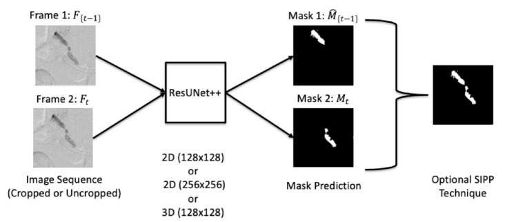
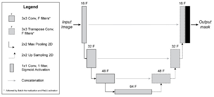
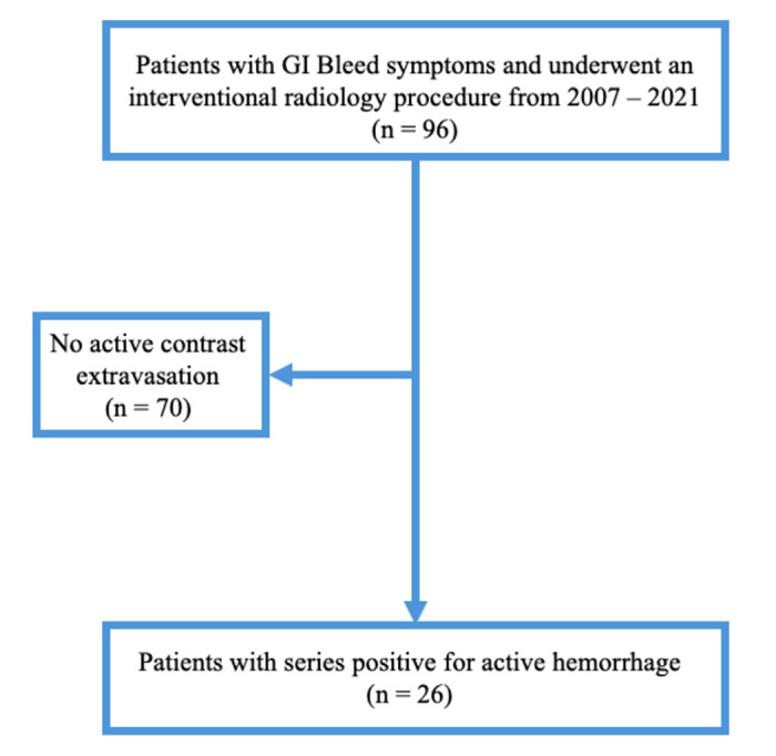
Methods: All mesenteric artery angiograms and arterial embolization DSA images obtained in the interventional radiology department between January 1, 2007, and December 31, 2021, were analyzed. These images were acquired using fluoroscopy imaging systems (Siemens Healthineers, USA). Thirty-nine unique series of bleeding images were augmented to train two-dimensional (2D) and three-dimensional (3D) residual neural networks (ResUNet++) for image segmentation. The 2D ResUNet++ network was trained on 3,548 images and tested on 394 images, whereas the 3D ResUNet++ network was trained on 316 3D objects and tested on 35 objects. For each case, both manually cropped images focused on the GI bleed and uncropped images were evaluated, with a superimposition post-processing (SIPP) technique applied to both image types.
Results: Based on both quantitative and qualitative analyses, the 2D ResUNet++ network significantly outperformed the 3D ResUNet++ model. In the qualitative evaluation, the 2D ResUNet++ model achieved the highest accuracy across both 128 × 128 and 256 × 256 input resolutions when enhanced with the SIPP technique, reaching accuracy rates between 95% and 97%. However, despite the improved detection consistency provided by SIPP, a reduction in Dice similarity coefficients was observed compared with models without post-processing. Specifically, the 2D ResUNet++ model combined with SIPP achieved a Dice accuracy of only 80%. This decline is primarily attributed to an increase in false positive predictions introduced by the temporal propagation of segmentation masks across frames.
Conclusion: Both 2D and 3D ResUNet++ networks can be trained to locate GI bleeding on DSA images prior to transarterial embolization. However, further research and refinement are needed before this technology can be implemented in DSA for real-time prediction.
Clinical significance: Automated detection of GI bleeding in DSA may reduce time to embolization, thereby improving patient outcomes.
期刊介绍:
Diagnostic and Interventional Radiology (Diagn Interv Radiol) is the open access, online-only official publication of Turkish Society of Radiology. It is published bimonthly and the journal’s publication language is English.
The journal is a medium for original articles, reviews, pictorial essays, technical notes related to all fields of diagnostic and interventional radiology.

 求助内容:
求助内容: 应助结果提醒方式:


