Differentiating oncocytic carcinoma from oncocytic adenoma: a comprehensive evaluation of preoperative characteristics and diagnostic approaches in a retrospective cohort study.
Eunji Kim, Jun Hyun Park, Ji-Young Park, Sang-Woo Lee, Jin Hyang Jung
{"title":"Differentiating oncocytic carcinoma from oncocytic adenoma: a comprehensive evaluation of preoperative characteristics and diagnostic approaches in a retrospective cohort study.","authors":"Eunji Kim, Jun Hyun Park, Ji-Young Park, Sang-Woo Lee, Jin Hyang Jung","doi":"10.4174/astr.2025.109.2.105","DOIUrl":null,"url":null,"abstract":"<p><strong>Purpose: </strong>Oncocytic carcinoma (OCA) has been reclassified from follicular thyroid carcinoma due to its unique features. Its rarity has resulted in limited studies on differentiating OCA from oncocytic adenoma (OA). This study aimed to compare the clinicopathologic and preoperative features of OCA and OA and evaluate the effectiveness of ultrasonography and cytology.</p><p><strong>Methods: </strong>We conducted a retrospective study involving 83 patients (23 with OCA and 60 with OA) who underwent thyroid surgery between 2011 and 2024. We reviewed clinical, ultrasonographic, cytologic, and histopathologic data to assess diagnostic performance.</p><p><strong>Results: </strong>OCA cases had larger tumors than OA in both sonographic (4.2 ± 1.7 cm <i>vs.</i> 2.7 ± 1.4 cm, P < 0.001) and pathologic measurements (3.8 ± 1.7 cm <i>vs.</i> 2.3 ± 1.4 cm, P < 0.001). K-TIRADS (the Korean Thyroid Imaging Reporting and Data System) did not effectively distinguish OCA from OA; however, ACR TI-RADS (the American College of Radiology Thyroid Imaging Reporting and Data System) categorized more OCA cases into higher-risk groups (17.4% <i>vs.</i> 1.7%, P = 0.016). Trabecular formation and intranodular vascularity were more frequent in OCA (17.4% <i>vs.</i> 1.7%, P = 0.019; 65.2% <i>vs.</i> 33.3%, P = 0.049). Cytologically, 87% of OCAs were classified as follicular neoplasms compared to 20% of OAs.</p><p><strong>Conclusion: </strong>Predicting malignancy in oncocytic neoplasms is challenging. Larger tumor size, higher ACR TI-RADS scores, and trabecular formation are potential indicators for OCA. Cytologic subcategorization within Bethesda IV suggests follicular neoplasms carry a higher malignancy risk than oncocytic neoplasms. Multicenter studies are needed to validate these findings.</p>","PeriodicalId":8071,"journal":{"name":"Annals of Surgical Treatment and Research","volume":"109 2","pages":"105-112"},"PeriodicalIF":1.6000,"publicationDate":"2025-08-01","publicationTypes":"Journal Article","fieldsOfStudy":null,"isOpenAccess":false,"openAccessPdf":"https://www.ncbi.nlm.nih.gov/pmc/articles/PMC12329133/pdf/","citationCount":"0","resultStr":null,"platform":"Semanticscholar","paperid":null,"PeriodicalName":"Annals of Surgical Treatment and Research","FirstCategoryId":"3","ListUrlMain":"https://doi.org/10.4174/astr.2025.109.2.105","RegionNum":4,"RegionCategory":"医学","ArticlePicture":[],"TitleCN":null,"AbstractTextCN":null,"PMCID":null,"EPubDate":"2025/7/30 0:00:00","PubModel":"Epub","JCR":"Q3","JCRName":"SURGERY","Score":null,"Total":0}
引用次数: 0
Abstract
Purpose: Oncocytic carcinoma (OCA) has been reclassified from follicular thyroid carcinoma due to its unique features. Its rarity has resulted in limited studies on differentiating OCA from oncocytic adenoma (OA). This study aimed to compare the clinicopathologic and preoperative features of OCA and OA and evaluate the effectiveness of ultrasonography and cytology.
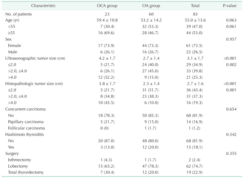
Methods: We conducted a retrospective study involving 83 patients (23 with OCA and 60 with OA) who underwent thyroid surgery between 2011 and 2024. We reviewed clinical, ultrasonographic, cytologic, and histopathologic data to assess diagnostic performance.
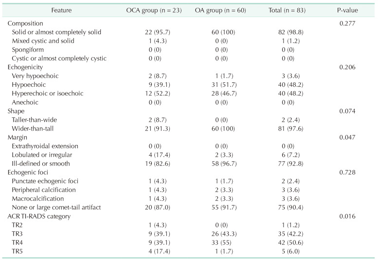
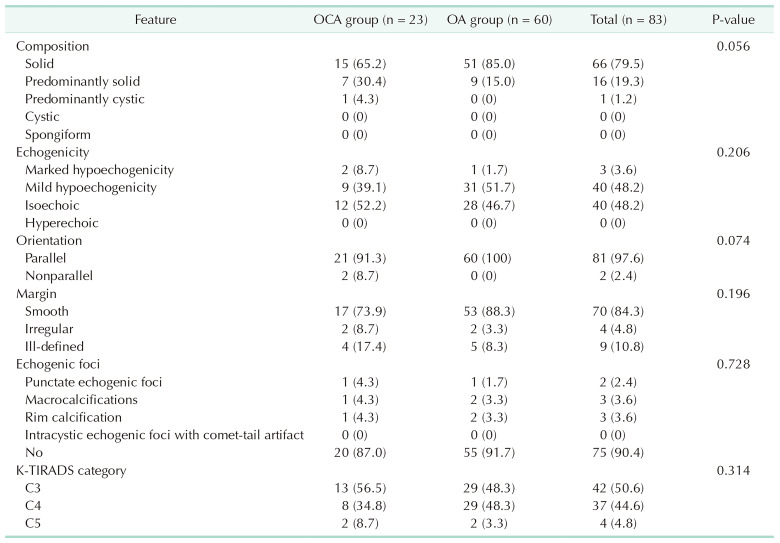
Results: OCA cases had larger tumors than OA in both sonographic (4.2 ± 1.7 cm vs. 2.7 ± 1.4 cm, P < 0.001) and pathologic measurements (3.8 ± 1.7 cm vs. 2.3 ± 1.4 cm, P < 0.001). K-TIRADS (the Korean Thyroid Imaging Reporting and Data System) did not effectively distinguish OCA from OA; however, ACR TI-RADS (the American College of Radiology Thyroid Imaging Reporting and Data System) categorized more OCA cases into higher-risk groups (17.4% vs. 1.7%, P = 0.016). Trabecular formation and intranodular vascularity were more frequent in OCA (17.4% vs. 1.7%, P = 0.019; 65.2% vs. 33.3%, P = 0.049). Cytologically, 87% of OCAs were classified as follicular neoplasms compared to 20% of OAs.
Conclusion: Predicting malignancy in oncocytic neoplasms is challenging. Larger tumor size, higher ACR TI-RADS scores, and trabecular formation are potential indicators for OCA. Cytologic subcategorization within Bethesda IV suggests follicular neoplasms carry a higher malignancy risk than oncocytic neoplasms. Multicenter studies are needed to validate these findings.
目的:嗜瘤细胞癌(OCA)因其独特的特征而被重新分类为滤泡性甲状腺癌。由于其罕见性,导致对OCA与癌细胞性腺瘤(OA)鉴别的研究有限。本研究旨在比较OCA和OA的临床病理和术前特征,并评价超声和细胞学检查的有效性。方法:我们对2011年至2024年间接受甲状腺手术的83例患者(23例OCA, 60例OA)进行了回顾性研究。我们回顾了临床、超声、细胞学和组织病理学资料来评估诊断表现。结果:超声(4.2±1.7 cm vs. 2.7±1.4 cm, P < 0.001)和病理(3.8±1.7 cm vs. 2.3±1.4 cm, P < 0.001)显示OCA患者肿瘤大于OA。K-TIRADS(韩国甲状腺成像报告和数据系统)不能有效区分OCA和OA;然而,ACR TI-RADS(美国放射学会甲状腺影像学报告和数据系统)将更多的OCA病例分类为高风险组(17.4%比1.7%,P = 0.016)。小梁形成和结节内血管在OCA中更为常见(17.4% vs. 1.7%, P = 0.019;65.2% vs. 33.3%, P = 0.049)。细胞学上,87%的oca被归类为滤泡性肿瘤,而20%的oa被归类为滤泡性肿瘤。结论:预测嗜瘤细胞肿瘤的恶性是具有挑战性的。较大的肿瘤大小、较高的ACR TI-RADS评分和小梁形成是OCA的潜在指标。Bethesda IV的细胞学亚分类表明,滤泡性肿瘤比嗜酸细胞性肿瘤具有更高的恶性风险。需要多中心研究来验证这些发现。
期刊介绍:
Manuscripts to the Annals of Surgical Treatment and Research (Ann Surg Treat Res) should be written in English according to the instructions for authors. If the details are not described below, the style should follow the Uniform Requirements for Manuscripts Submitted to Biomedical Journals: Writing and Editing for Biomedical Publications available at International Committee of Medical Journal Editors (ICMJE) website (http://www.icmje.org).

 求助内容:
求助内容: 应助结果提醒方式:


