Altered white matter integrity and structural network topology in rhegmatogenous retinal detachment: A diffusion tensor imaging study
IF 2.6
4区 医学
Q3 NEUROSCIENCES
引用次数: 0
Abstract
Background
Rhegmatogenous retinal detachment (RRD) has been associated with gray matter alterations, but its effects on white matter microstructure and brain network organization remain largely unexplored.
Methods
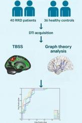
This study included 40 RRD patients and 36 healthy controls (HCs), who underwent diffusion tensor imaging (DTI). Tract-Based Spatial Statistics (TBSS) was used to assess white matter microstructure, and graph theory was applied to quantify structural network topology. In addition, a support vector machine (SVM) classifier was trained to evaluate the discriminative potential of imaging-derived features.
Results
Compared to HCs, RRD patients exhibited disrupted white matter network topology, characterized by reduced small-world properties and increased global efficiency. Regionally, widespread alterations in nodal centrality and efficiency were observed, primarily in the frontal, temporal, and occipital lobes. Structural connectivity analysis revealed enhanced integration between attention-related networks and diminished within-network coherence in the default mode and dorsal attention systems. TBSS further identified microstructural abnormalities in the corpus callosum and corona radiata. Notably, degree centrality (DC) achieved the highest classification accuracy in SVM, with an area under the curve (AUC) of 0.9125.
Conclusion
RRD patients exhibit widespread alterations in white matter microstructure and structural network topology, indicating central nervous system involvement following acute peripheral visual loss. Among network metrics, DC showed the highest discriminative power. These findings offer preliminary insights into the neural mechanisms of RRD and may inform future studies on disease stratification or prognosis.
孔源性视网膜脱离的白质完整性和结构网络拓扑改变:扩散张量成像研究。
背景:孔源性视网膜脱离(RRD)与灰质改变有关,但其对白质微观结构和脑网络组织的影响仍未被广泛研究。方法:本研究纳入40例RRD患者和36例健康对照(hc),均行弥散张量成像(DTI)检查。利用基于tractbased Spatial Statistics (TBSS)对白质微观结构进行评估,利用图论对结构网络拓扑进行量化。此外,还训练了支持向量机(SVM)分类器来评估图像衍生特征的判别潜力。结果:与hc相比,RRD患者表现出白质网络拓扑结构紊乱,其特征是小世界特性降低,整体效率提高。在区域上,观察到淋巴结中心性和效率的广泛改变,主要发生在额叶、颞叶和枕叶。结构连通性分析表明,默认模式和背侧注意系统的注意相关网络之间的整合增强,网络内一致性减弱。TBSS进一步发现胼胝体和辐射冠的显微结构异常。值得注意的是,度中心性(DC)的分类准确率最高,曲线下面积(AUC)为0.9125。结论:RRD患者表现出广泛的白质微观结构和结构网络拓扑改变,表明急性周围视力丧失后中枢神经系统受损伤。在网络指标中,DC表现出最高的判别能力。这些发现为RRD的神经机制提供了初步的见解,并可能为未来的疾病分层或预后研究提供信息。
本文章由计算机程序翻译,如有差异,请以英文原文为准。
求助全文
约1分钟内获得全文
求助全文
来源期刊
Brain Research
医学-神经科学
CiteScore
5.90
自引率
3.40%
发文量
268
审稿时长
47 days
期刊介绍:
An international multidisciplinary journal devoted to fundamental research in the brain sciences.
Brain Research publishes papers reporting interdisciplinary investigations of nervous system structure and function that are of general interest to the international community of neuroscientists. As is evident from the journals name, its scope is broad, ranging from cellular and molecular studies through systems neuroscience, cognition and disease. Invited reviews are also published; suggestions for and inquiries about potential reviews are welcomed.
With the appearance of the final issue of the 2011 subscription, Vol. 67/1-2 (24 June 2011), Brain Research Reviews has ceased publication as a distinct journal separate from Brain Research. Review articles accepted for Brain Research are now published in that journal.
 求助内容:
求助内容: 应助结果提醒方式:


