Neuronal Colocalization of μ-Opioid Receptor, κ-Opioid Receptor, and Oxytocin Receptor mRNA in the Central Nucleus of the Amygdala in Male and Female Mice.
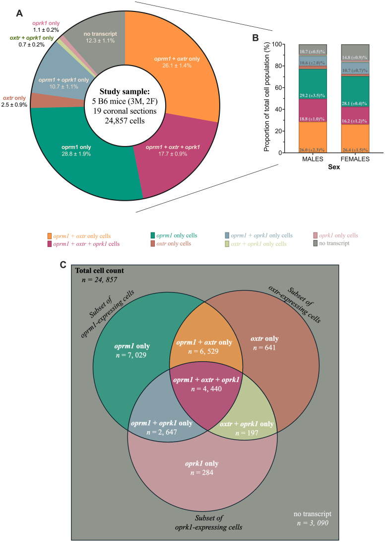
{"title":"Neuronal Colocalization of μ-Opioid Receptor, κ-Opioid Receptor, and Oxytocin Receptor mRNA in the Central Nucleus of the Amygdala in Male and Female Mice.","authors":"Khalin E Nisbett, George F Koob","doi":"10.1523/ENEURO.0059-25.2025","DOIUrl":null,"url":null,"abstract":"<p><p>Given the observed interaction and reports of oxytocin, μ-opioid receptor, or κ-opioid receptor expression in brain regions important to emotion regulation (i.e., the central amygdala), we hypothesized that oxytocin (<i>oxtr</i>), μ-opioid (<i>oprm1</i>), and κ-opioid (<i>oprk1</i>) receptor mRNA were colocalized to the same cells in the central amygdala. RNAscope in situ hybridization performed on fresh-frozen coronal brain sections was used to label cells containing <i>oxtr</i>, <i>oprm1</i>, and/or <i>oprk1</i> The coronal sections were imaged using a 40× objective (widefield fluorescence) on a Leica Thunder fluorescent microscope, and the images were processed using open-source ImageJ/Fiji software and analyzed using the Imaris software. The central amygdala was identified using Paxinos and Watson's <i>The Mouse Brain in Stereotaxic Coordinates</i> ( Paxinos and Franklin, 2019). Eight distinct cell populations were enumerated (i.e., <i>oxtr</i>-only, <i>oprm1</i>-only, <i>oprk1</i>-only, <i>oxtr</i> + <i>oprm1</i>-only, <i>oxtr</i> + <i>oprk1</i>-only, <i>oprm1</i> + <i>oprk1</i>-only, <i>oxtr</i> + <i>oprm1</i> + <i>oprk1</i>, and nontranscript cells). Our findings demonstrated that 47% of cells in the central amygdala express <i>oxtr</i> with <i>oprm1</i> and/or <i>oprk1</i> Of the <i>oxtr</i>-expressing cells, 38% colocalized only <i>oprm1</i>, and 56% of <i>oxtr</i>-expressing cells colocalized both <i>oprm1</i> and <i>oprk1</i> However, 53% of <i>oprm1</i>-expressing cells colocalized <i>oxtr</i>, and 61% of <i>oprk1</i>-expressiong cells colocalized <i>oxtr</i> These findings suggest that opioid and oxytocin receptors can function at the cellular level through morphological interactions. Future work will examine the physiological basis for the interaction between opioid and oxytocin receptors using transgenic behavior and electrophysiological assays.</p>","PeriodicalId":11617,"journal":{"name":"eNeuro","volume":" ","pages":""},"PeriodicalIF":2.7000,"publicationDate":"2025-09-10","publicationTypes":"Journal Article","fieldsOfStudy":null,"isOpenAccess":false,"openAccessPdf":"https://www.ncbi.nlm.nih.gov/pmc/articles/PMC12439755/pdf/","citationCount":"0","resultStr":null,"platform":"Semanticscholar","paperid":null,"PeriodicalName":"eNeuro","FirstCategoryId":"3","ListUrlMain":"https://doi.org/10.1523/ENEURO.0059-25.2025","RegionNum":3,"RegionCategory":"医学","ArticlePicture":[],"TitleCN":null,"AbstractTextCN":null,"PMCID":null,"EPubDate":"2025/9/1 0:00:00","PubModel":"Print","JCR":"Q3","JCRName":"NEUROSCIENCES","Score":null,"Total":0}
引用次数: 0
Abstract
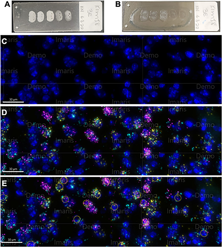
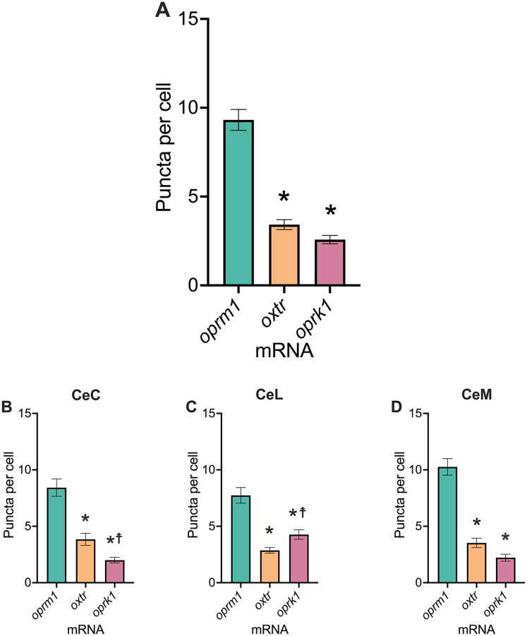
Given the observed interaction and reports of oxytocin, μ-opioid receptor, or κ-opioid receptor expression in brain regions important to emotion regulation (i.e., the central amygdala), we hypothesized that oxytocin (oxtr), μ-opioid (oprm1), and κ-opioid (oprk1) receptor mRNA were colocalized to the same cells in the central amygdala. RNAscope in situ hybridization performed on fresh-frozen coronal brain sections was used to label cells containing oxtr, oprm1, and/or oprk1 The coronal sections were imaged using a 40× objective (widefield fluorescence) on a Leica Thunder fluorescent microscope, and the images were processed using open-source ImageJ/Fiji software and analyzed using the Imaris software. The central amygdala was identified using Paxinos and Watson's The Mouse Brain in Stereotaxic Coordinates ( Paxinos and Franklin, 2019). Eight distinct cell populations were enumerated (i.e., oxtr-only, oprm1-only, oprk1-only, oxtr + oprm1-only, oxtr + oprk1-only, oprm1 + oprk1-only, oxtr + oprm1 + oprk1, and nontranscript cells). Our findings demonstrated that 47% of cells in the central amygdala express oxtr with oprm1 and/or oprk1 Of the oxtr-expressing cells, 38% colocalized only oprm1, and 56% of oxtr-expressing cells colocalized both oprm1 and oprk1 However, 53% of oprm1-expressing cells colocalized oxtr, and 61% of oprk1-expressiong cells colocalized oxtr These findings suggest that opioid and oxytocin receptors can function at the cellular level through morphological interactions. Future work will examine the physiological basis for the interaction between opioid and oxytocin receptors using transgenic behavior and electrophysiological assays.
期刊介绍:
An open-access journal from the Society for Neuroscience, eNeuro publishes high-quality, broad-based, peer-reviewed research focused solely on the field of neuroscience. eNeuro embodies an emerging scientific vision that offers a new experience for authors and readers, all in support of the Society’s mission to advance understanding of the brain and nervous system.

 求助内容:
求助内容: 应助结果提醒方式:


