{"title":"Coptisine alleviates high glucose-induced HUVEC dysfunction in vitro and inhibits gestational diabetes mellitus in vivo","authors":"Lan Wang, Wei Xiong, Yunyun Jiang","doi":"10.1007/s10735-025-10542-z","DOIUrl":null,"url":null,"abstract":"<div><p>The dysfunction of vascular endothelial cells (VECs) is an important cause of diabetes-related cardiovascular diseases. Coptisine is a bioactive component of <i>Rhizoma coptidis</i> with anti-diabetes property. The aim of this present study was to clarify the role and possible mechanism of coptisine underlying VEC dysfunction during gestational diabetes mellitus (GDM). Human umbilical vein endothelial cells (HUVECs) were treated with coptisine and/or high glucose (HG) medium. A GDM rat model was established by high-fat diet feeding and streptozotocin injection. Cell viability, migration, and angiogenesis were detected by 3-(4,5-dimethylthiazol-2-yl)-2,5-diphenyltetrazolium bromide assay, wound healing assay, Transwell assay, and tube formation assay. The levels of angiogenesis-related proteins and the key markers in the Adenosine monophosphateactivated protein kinase (AMPK)/nuclear factor-erythroid 2-related factor 2 (NRF2) signaling pathway were tested by western blot analysis. The pathological changes of placental tissues were observed by HE staining. We found that the cell viability of HUVECs was repressed in HG conditions in a dose-dependent manner, and 25 mM HG reduced the HUVEC viability in a time-dependent manner. Coptisine (5 to 5o μM) did not cause cytotoxicity to HUVECs. In addition, HG-induced the decrease in cell viability and migration of HUVECs were rescued by coptisine in a dose-dependent manner. Coptisine restored the angiogenetic ability of HG-induced HUVECs by upregulating the protein expression of fibroblast growth factor 2, vascular endothelial-derived growth factor, and Angiotensin 1. Moreover, coptisine treatment re-activated the AMPK/NRF2 pathway in HG-stimulated HUVECs. Importantly, inhibition of AMPK/NRF2 signaling reverses the effect of coptisine on cell viability, migration, and angiogenesis of HUVECs. The in vivo study demonstrated that coptisine suppresses hyperglycemia and placenta injury in GDM rats. Coptisine protected HUVECs from hyperglycemic insult, suggesting the potential of coptisine which might be used as a therapeutic agent for VEC dysfunction in GDM.</p></div>","PeriodicalId":650,"journal":{"name":"Journal of Molecular Histology","volume":"56 4","pages":""},"PeriodicalIF":2.2000,"publicationDate":"2025-08-09","publicationTypes":"Journal Article","fieldsOfStudy":null,"isOpenAccess":false,"openAccessPdf":"","citationCount":"0","resultStr":null,"platform":"Semanticscholar","paperid":null,"PeriodicalName":"Journal of Molecular Histology","FirstCategoryId":"99","ListUrlMain":"https://link.springer.com/article/10.1007/s10735-025-10542-z","RegionNum":4,"RegionCategory":"生物学","ArticlePicture":[],"TitleCN":null,"AbstractTextCN":null,"PMCID":null,"EPubDate":"","PubModel":"","JCR":"Q3","JCRName":"CELL BIOLOGY","Score":null,"Total":0}
引用次数: 0
Abstract
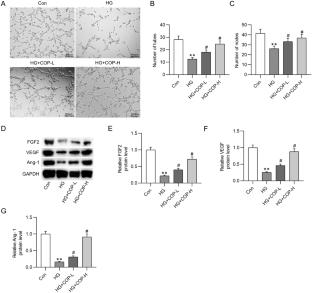
The dysfunction of vascular endothelial cells (VECs) is an important cause of diabetes-related cardiovascular diseases. Coptisine is a bioactive component of Rhizoma coptidis with anti-diabetes property. The aim of this present study was to clarify the role and possible mechanism of coptisine underlying VEC dysfunction during gestational diabetes mellitus (GDM). Human umbilical vein endothelial cells (HUVECs) were treated with coptisine and/or high glucose (HG) medium. A GDM rat model was established by high-fat diet feeding and streptozotocin injection. Cell viability, migration, and angiogenesis were detected by 3-(4,5-dimethylthiazol-2-yl)-2,5-diphenyltetrazolium bromide assay, wound healing assay, Transwell assay, and tube formation assay. The levels of angiogenesis-related proteins and the key markers in the Adenosine monophosphateactivated protein kinase (AMPK)/nuclear factor-erythroid 2-related factor 2 (NRF2) signaling pathway were tested by western blot analysis. The pathological changes of placental tissues were observed by HE staining. We found that the cell viability of HUVECs was repressed in HG conditions in a dose-dependent manner, and 25 mM HG reduced the HUVEC viability in a time-dependent manner. Coptisine (5 to 5o μM) did not cause cytotoxicity to HUVECs. In addition, HG-induced the decrease in cell viability and migration of HUVECs were rescued by coptisine in a dose-dependent manner. Coptisine restored the angiogenetic ability of HG-induced HUVECs by upregulating the protein expression of fibroblast growth factor 2, vascular endothelial-derived growth factor, and Angiotensin 1. Moreover, coptisine treatment re-activated the AMPK/NRF2 pathway in HG-stimulated HUVECs. Importantly, inhibition of AMPK/NRF2 signaling reverses the effect of coptisine on cell viability, migration, and angiogenesis of HUVECs. The in vivo study demonstrated that coptisine suppresses hyperglycemia and placenta injury in GDM rats. Coptisine protected HUVECs from hyperglycemic insult, suggesting the potential of coptisine which might be used as a therapeutic agent for VEC dysfunction in GDM.
期刊介绍:
The Journal of Molecular Histology publishes results of original research on the localization and expression of molecules in animal cells, tissues and organs. Coverage includes studies describing novel cellular or ultrastructural distributions of molecules which provide insight into biochemical or physiological function, development, histologic structure and disease processes.
Major research themes of particular interest include:
- Cell-Cell and Cell-Matrix Interactions;
- Connective Tissues;
- Development and Disease;
- Neuroscience.
Please note that the Journal of Molecular Histology does not consider manuscripts dealing with the application of immunological or other probes on non-standard laboratory animal models unless the results are clearly of significant and general biological importance.
The Journal of Molecular Histology publishes full-length original research papers, review articles, short communications and letters to the editors. All manuscripts are typically reviewed by two independent referees. The Journal of Molecular Histology is a continuation of The Histochemical Journal.

 求助内容:
求助内容: 应助结果提醒方式:


