Vittorio Patanè, Umberto Atripaldi, Mario Sansone, Luca Marinelli, Sara Del Tufo, Gianluca Arrichiello, Davide Ciardiello, Francesco Selvaggi, Erika Martinelli, Alfonso Reginelli
{"title":"MRI-based radiomics for preoperative T-staging of rectal cancer: a retrospective analysis.","authors":"Vittorio Patanè, Umberto Atripaldi, Mario Sansone, Luca Marinelli, Sara Del Tufo, Gianluca Arrichiello, Davide Ciardiello, Francesco Selvaggi, Erika Martinelli, Alfonso Reginelli","doi":"10.1007/s00384-025-04969-9","DOIUrl":null,"url":null,"abstract":"<p><strong>Puropose: </strong>Preoperative T-staging in rectal cancer is essential for treatment planning, yet conventional MRI shows limited accuracy (~ 60-78). Our study investigates whether radiomic analysis of high-resolution T2-weighted MRI can non-invasively improve staging accuracy through a retrospective evaluation in a real-world surgical cohort.</p><p><strong>Methods: </strong>This single-center retrospective study included 200 patients (January 2024-April 2025) with pathologically confirmed rectal cancer, all undergoing preoperative high-resolution T2-weighted MRI within one week prior to curative surgery and no neoadjuvant therapy. Manual segmentation was performed using ITK‑SNAP, followed by extraction of 107 radiomic features via PyRadiomics. Feature selection employed mRMR and LASSO logistic regression, culminating in a Rad-score predictive model. Statistical performance was evaluated using ROC curves (AUC), accuracy, sensitivity, specificity, and Delong's test.</p><p><strong>Results: </strong>Among 200 patients, 95 were pathologically staged as T2 and 105 as T3-T4 (55 T3, 50 T4). After preprocessing, 26 radiomic features were retained; key features including ngtdm_contrast and ngtdm_coarseness showed AUC values > 0.70. The LASSO-based model achieved an AUC of 0.82 (95% CI: 0.75-0.89), with overall accuracy of 81%, sensitivity of 78%, and specificity of 84%.</p><p><strong>Conclusion: </strong>Radiomic analysis of standard preoperative T2-weighted MRI provides a reliable, non-invasive method to predict rectal cancer T-stage. This approach has the potential to enhance staging accuracy and inform personalized surgical planning. Prospective multicenter validation is required for broader clinical implementation.</p>","PeriodicalId":13789,"journal":{"name":"International Journal of Colorectal Disease","volume":"40 1","pages":"174"},"PeriodicalIF":2.3000,"publicationDate":"2025-08-08","publicationTypes":"Journal Article","fieldsOfStudy":null,"isOpenAccess":false,"openAccessPdf":"https://www.ncbi.nlm.nih.gov/pmc/articles/PMC12334464/pdf/","citationCount":"0","resultStr":null,"platform":"Semanticscholar","paperid":null,"PeriodicalName":"International Journal of Colorectal Disease","FirstCategoryId":"3","ListUrlMain":"https://doi.org/10.1007/s00384-025-04969-9","RegionNum":3,"RegionCategory":"医学","ArticlePicture":[],"TitleCN":null,"AbstractTextCN":null,"PMCID":null,"EPubDate":"","PubModel":"","JCR":"Q2","JCRName":"GASTROENTEROLOGY & HEPATOLOGY","Score":null,"Total":0}
引用次数: 0
Abstract
Puropose: Preoperative T-staging in rectal cancer is essential for treatment planning, yet conventional MRI shows limited accuracy (~ 60-78). Our study investigates whether radiomic analysis of high-resolution T2-weighted MRI can non-invasively improve staging accuracy through a retrospective evaluation in a real-world surgical cohort.
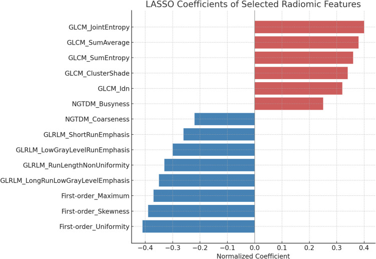
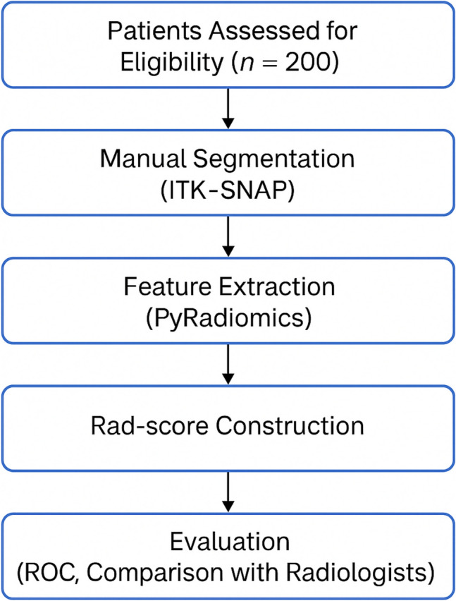
Methods: This single-center retrospective study included 200 patients (January 2024-April 2025) with pathologically confirmed rectal cancer, all undergoing preoperative high-resolution T2-weighted MRI within one week prior to curative surgery and no neoadjuvant therapy. Manual segmentation was performed using ITK‑SNAP, followed by extraction of 107 radiomic features via PyRadiomics. Feature selection employed mRMR and LASSO logistic regression, culminating in a Rad-score predictive model. Statistical performance was evaluated using ROC curves (AUC), accuracy, sensitivity, specificity, and Delong's test.
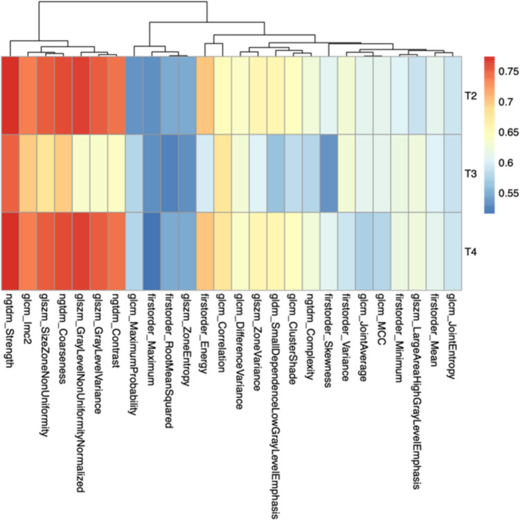
Results: Among 200 patients, 95 were pathologically staged as T2 and 105 as T3-T4 (55 T3, 50 T4). After preprocessing, 26 radiomic features were retained; key features including ngtdm_contrast and ngtdm_coarseness showed AUC values > 0.70. The LASSO-based model achieved an AUC of 0.82 (95% CI: 0.75-0.89), with overall accuracy of 81%, sensitivity of 78%, and specificity of 84%.
Conclusion: Radiomic analysis of standard preoperative T2-weighted MRI provides a reliable, non-invasive method to predict rectal cancer T-stage. This approach has the potential to enhance staging accuracy and inform personalized surgical planning. Prospective multicenter validation is required for broader clinical implementation.
期刊介绍:
The International Journal of Colorectal Disease, Clinical and Molecular Gastroenterology and Surgery aims to publish novel and state-of-the-art papers which deal with the physiology and pathophysiology of diseases involving the entire gastrointestinal tract. In addition to original research articles, the following categories will be included: reviews (usually commissioned but may also be submitted), case reports, letters to the editor, and protocols on clinical studies.
The journal offers its readers an interdisciplinary forum for clinical science and molecular research related to gastrointestinal disease.
 求助内容:
求助内容: 应助结果提醒方式:


