A Case of False-Negative Results in the One-Step Nucleic Acid Amplification Assay for Sentinel Lymph Node Metastasis in Breast Cancer with Low Cytokeratin 19 Expression.
Ai Koyanagi, Akinari Kakumoto, Hajime Kuroda, Shogo Baba, Mami Koketsu, Oi Harada, Yasutaka Kato, Hiroshi Nishihara, Hiroyuki Kawami
{"title":"A Case of False-Negative Results in the One-Step Nucleic Acid Amplification Assay for Sentinel Lymph Node Metastasis in Breast Cancer with Low Cytokeratin 19 Expression.","authors":"Ai Koyanagi, Akinari Kakumoto, Hajime Kuroda, Shogo Baba, Mami Koketsu, Oi Harada, Yasutaka Kato, Hiroshi Nishihara, Hiroyuki Kawami","doi":"10.1159/000546995","DOIUrl":null,"url":null,"abstract":"<p><strong>Introduction: </strong>One-step nucleic acid amplification (OSNA) for the analysis of sentinel lymph nodes is now widely used as a reliable tool for the intraoperative diagnosis of breast cancer metastasis based on the quantification of CK19 mRNA. However, discrepancies have been noted between the molecular diagnosis and histological evaluation, potentially due to differences in tissue sampling or technical limitations. Furthermore, false-negative results may occur when target mRNA expression is reduced.</p><p><strong>Case presentation: </strong>We herein describe a 45-year-old female patient who underwent breast-conserving surgery. The final pathological stage after surgery was pT2N1aM0 (stage IIB), hormone receptor positive (HER2: 1+). An OSNA analysis of two sentinel lymph nodes revealed no metastasis, whereas touch imprint cytology of one of the lymph nodes was positive. Immunohistochemistry of the primary tumor showed a mixture of extensive CK19-negative and focal CK19-positive lesions. A genetic analysis of the CK19-negative lesion detected mutations and abnormalities, including the <i>ESR1</i> amplification, <i>FANCA</i> deletion, <i>STK11</i> deletion, <i>TSC2</i> deletion, <i>POLD1</i> deletion, and mutations in <i>PIK3CA</i>, <i>ARID1A</i>, <i>NF2</i>, <i>SETBP1</i>, and <i>EP300</i>. Of these, tumor heterogeneity and genetic alterations, including mutations in <i>ARID1A</i>, <i>NF2</i>, and <i>EP300</i>, in the CK19-negative lesion were hypothesized to suppress CK19 expression.</p><p><strong>Conclusion: </strong>False-negative results are more likely to occur for CK19-negative breast cancer, emphasizing the importance of incorporating complementary diagnostic methods, such as touch imprint cytology and different molecular markers. A multifaceted diagnostic approach is crucial for ensuring accurate staging and appropriate treatment planning.</p>","PeriodicalId":9625,"journal":{"name":"Case Reports in Oncology","volume":"18 1","pages":"1074-1080"},"PeriodicalIF":0.7000,"publicationDate":"2025-07-02","publicationTypes":"Journal Article","fieldsOfStudy":null,"isOpenAccess":false,"openAccessPdf":"https://www.ncbi.nlm.nih.gov/pmc/articles/PMC12331226/pdf/","citationCount":"0","resultStr":null,"platform":"Semanticscholar","paperid":null,"PeriodicalName":"Case Reports in Oncology","FirstCategoryId":"1085","ListUrlMain":"https://doi.org/10.1159/000546995","RegionNum":0,"RegionCategory":null,"ArticlePicture":[],"TitleCN":null,"AbstractTextCN":null,"PMCID":null,"EPubDate":"2025/1/1 0:00:00","PubModel":"eCollection","JCR":"Q4","JCRName":"ONCOLOGY","Score":null,"Total":0}
引用次数: 0
Abstract
Introduction: One-step nucleic acid amplification (OSNA) for the analysis of sentinel lymph nodes is now widely used as a reliable tool for the intraoperative diagnosis of breast cancer metastasis based on the quantification of CK19 mRNA. However, discrepancies have been noted between the molecular diagnosis and histological evaluation, potentially due to differences in tissue sampling or technical limitations. Furthermore, false-negative results may occur when target mRNA expression is reduced.
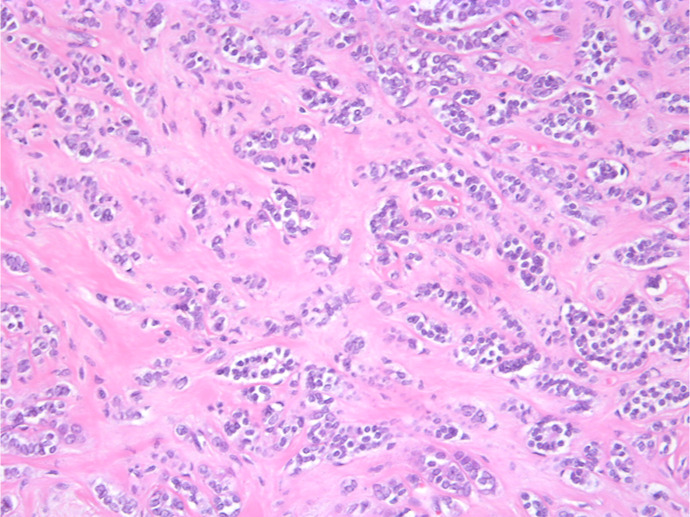
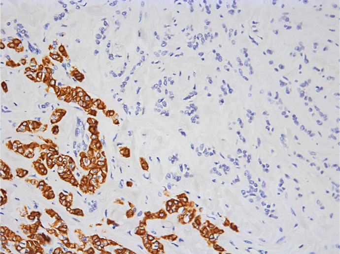
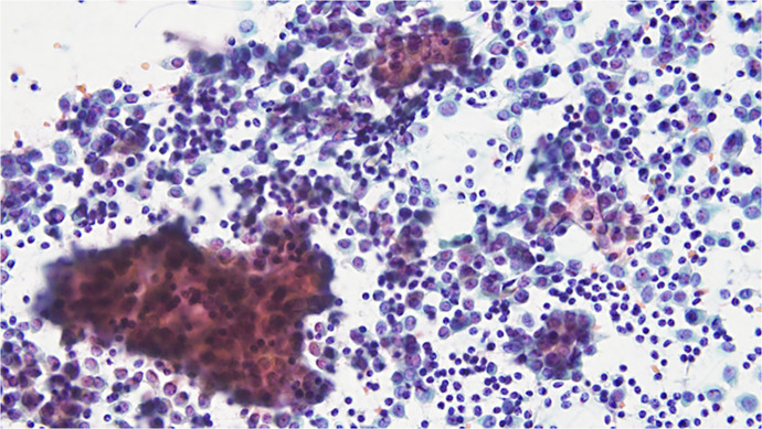
Case presentation: We herein describe a 45-year-old female patient who underwent breast-conserving surgery. The final pathological stage after surgery was pT2N1aM0 (stage IIB), hormone receptor positive (HER2: 1+). An OSNA analysis of two sentinel lymph nodes revealed no metastasis, whereas touch imprint cytology of one of the lymph nodes was positive. Immunohistochemistry of the primary tumor showed a mixture of extensive CK19-negative and focal CK19-positive lesions. A genetic analysis of the CK19-negative lesion detected mutations and abnormalities, including the ESR1 amplification, FANCA deletion, STK11 deletion, TSC2 deletion, POLD1 deletion, and mutations in PIK3CA, ARID1A, NF2, SETBP1, and EP300. Of these, tumor heterogeneity and genetic alterations, including mutations in ARID1A, NF2, and EP300, in the CK19-negative lesion were hypothesized to suppress CK19 expression.
Conclusion: False-negative results are more likely to occur for CK19-negative breast cancer, emphasizing the importance of incorporating complementary diagnostic methods, such as touch imprint cytology and different molecular markers. A multifaceted diagnostic approach is crucial for ensuring accurate staging and appropriate treatment planning.


 求助内容:
求助内容: 应助结果提醒方式:


