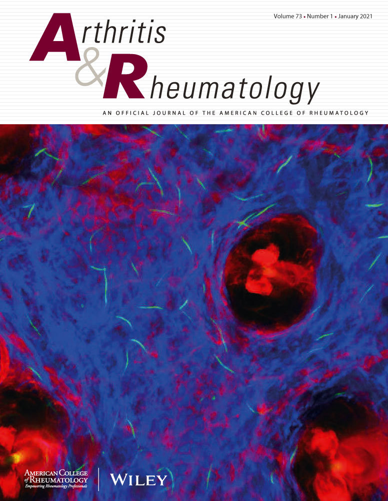
Epidermal Interferon kappa drives cutaneous lupus-like lesions, photosensitivity and systemic autoimmunity in vivo.
IF 10.9
1区 医学
Q1 RHEUMATOLOGY
引用次数: 0
Abstract
OBJECTIVE Keratinocyte-derived interferon kappa (IFN-κ) is chronically overexpressed in human non-lesional systemic lupus erythematosus (SLE) skin. Recent evidence suggests that epidermal signals instruct the immune system in SLE, but whether epidermal IFN-κ alone is sufficient to drive lupus phenotypes has not been investigated. This study aimed to identify whether epidermal-specific overexpression of Interferon kappa (Ifnk) results in lupus-like cutaneous and systemic inflammation. METHODS We compared three month (young) and twelve month (aged) old Balb/c mice that overexpress Ifnk in the epidermis under the keratin 14 promoter (transgenic, TG) with age-matched Balb/c wild type mice (WT) and assessed local and systemic immune responses at baseline and after UV exposure. Skin lesions were assessed by histopathology, bulk RNA sequencing and immunohistochemistry and subsequently compared to human cutaneous lupus erythematosus (CLE). Flow cytometry on lymph nodes and splenocytes at baseline and after UV exposure was performed to phenotype immune cell compositions. RESULTS Ifnk TG mice spontaneously develop CLE-like lesions and systemic immune dysregulation. Lesions show facial predominance, lymphocytic infiltration, immune complex deposition and a transcriptional signature reflective of human CLE. Ifnk TG mice exhibit increased immune cell activation and spontaneous signs of systemic autoimmunity with higher anti-dsDNA-antibodies, lymphadenopathy and splenomegaly, but lack signs of renal inflammation. UV exposure enhanced cutaneous inflammation and splenic T cell activation in Ifnk TG mice. CONCLUSION Together, we describe a new CLE mouse model that recapitulates features of human CLE and substantiates the role of epidermal IFN-κ as a driver of CLE, photosensitivity and systemic inflammation.表皮干扰素kappa驱动皮肤红斑狼疮样病变,光敏性和全身自身免疫。
目的:角化细胞源性干扰素κ (IFN-κ)在人非病变系统性红斑狼疮(SLE)皮肤中慢性过表达。最近的证据表明,表皮信号在SLE中指导免疫系统,但表皮IFN-κ是否足以单独驱动狼疮表型尚未研究。本研究旨在确定表皮特异性过表达干扰素kappa (Ifnk)是否会导致红斑狼疮样皮肤和全身炎症。方法我们比较了角蛋白14启动子(转基因,TG)下表皮过表达Ifnk的3月龄和12月龄Balb/c小鼠与年龄匹配的Balb/c野生型小鼠(WT),并评估了基线和紫外线照射后的局部和全身免疫反应。通过组织病理学、大量RNA测序和免疫组织化学评估皮肤病变,并随后与人类皮肤红斑狼疮(CLE)进行比较。在基线和紫外线照射后对淋巴结和脾细胞进行流式细胞术,以确定免疫细胞成分的表型。结果nk TG小鼠自发出现cle样病变和全身免疫失调。病变表现为面部优势,淋巴细胞浸润,免疫复合物沉积和反映人类CLE的转录特征。Ifnk TG小鼠表现出免疫细胞活化增加和自发的全身自身免疫征象(抗dsdna抗体升高)、淋巴结病和脾肿大,但缺乏肾脏炎症征象。紫外线暴露增强Ifnk TG小鼠皮肤炎症和脾T细胞活化。总之,我们描述了一个新的CLE小鼠模型,该模型概括了人类CLE的特征,并证实了表皮IFN-κ在CLE、光敏性和全身性炎症中的驱动作用。
本文章由计算机程序翻译,如有差异,请以英文原文为准。
求助全文
约1分钟内获得全文
求助全文
来源期刊

Arthritis & Rheumatology
RHEUMATOLOGY-
CiteScore
20.90
自引率
3.00%
发文量
371
期刊介绍:
Arthritis & Rheumatology is the official journal of the American College of Rheumatology and focuses on the natural history, pathophysiology, treatment, and outcome of rheumatic diseases. It is a peer-reviewed publication that aims to provide the highest quality basic and clinical research in this field. The journal covers a wide range of investigative areas and also includes review articles, editorials, and educational material for researchers and clinicians. Being recognized as a leading research journal in rheumatology, Arthritis & Rheumatology serves the global community of rheumatology investigators and clinicians.
 求助内容:
求助内容: 应助结果提醒方式:


