{"title":"Adrenal Incidentaloma Masquerading as Malignancy: A Rare Case of Ganglioneuroma with Nodal Metastasis.","authors":"Shahrukh Memon, Saurabh Raj, Rudrakshi Mahaldar","doi":"10.1159/000547075","DOIUrl":null,"url":null,"abstract":"<p><strong>Introduction: </strong>Adrenal ganglioneuromas are rare benign tumours, accounting for 0.3%-2% of adrenal incidentalomas. They arise from Schwann and ganglion cells and often mimic adrenal cortical carcinoma clinically and radiologically. Despite their benign nature, metastatic deposits in lymph nodes have been reported, suggesting tumour maturation. Diagnosis is typically confirmed through postoperative histopathology.</p><p><strong>Case presentation: </strong>A male patient in his early 60s with a history of hypertension presented with an adrenal incidentaloma. Imaging revealed a well-defined, hypodense right adrenal mass (35 HU) with calcifications and an enlarged aortocaval lymph node, raising suspicion for carcinoma. Biochemical analysis showed a non-functional adrenal tumour. Intraoperatively, the mass appeared benign, but the lymph node deposit suggested malignancy. Histopathological examination confirmed right adrenal ganglioneuroma with metastatic deposits in the aortocaval lymph nodes. The patient recovered well postoperatively, with normal serum cortisol levels and no signs of recurrence at 6-month follow-up.</p><p><strong>Conclusion: </strong>Adrenal ganglioneuromas can present as adrenal incidentalomas with lymph node involvement, mimicking malignancy. Surgery remains the gold standard for treatment, and postoperative histopathology is crucial for diagnosis. In such cases, no adjuvant therapy or stringent follow-up is required after surgical removal.</p>","PeriodicalId":9625,"journal":{"name":"Case Reports in Oncology","volume":"18 1","pages":"1057-1062"},"PeriodicalIF":0.7000,"publicationDate":"2025-06-25","publicationTypes":"Journal Article","fieldsOfStudy":null,"isOpenAccess":false,"openAccessPdf":"https://www.ncbi.nlm.nih.gov/pmc/articles/PMC12327930/pdf/","citationCount":"0","resultStr":null,"platform":"Semanticscholar","paperid":null,"PeriodicalName":"Case Reports in Oncology","FirstCategoryId":"1085","ListUrlMain":"https://doi.org/10.1159/000547075","RegionNum":0,"RegionCategory":null,"ArticlePicture":[],"TitleCN":null,"AbstractTextCN":null,"PMCID":null,"EPubDate":"2025/1/1 0:00:00","PubModel":"eCollection","JCR":"Q4","JCRName":"ONCOLOGY","Score":null,"Total":0}
引用次数: 0
Abstract
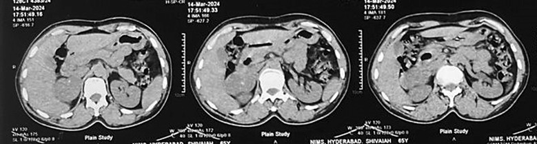
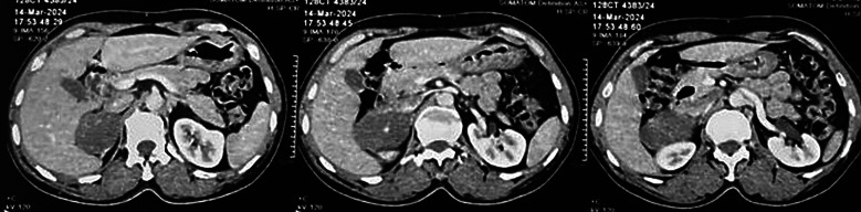
Introduction: Adrenal ganglioneuromas are rare benign tumours, accounting for 0.3%-2% of adrenal incidentalomas. They arise from Schwann and ganglion cells and often mimic adrenal cortical carcinoma clinically and radiologically. Despite their benign nature, metastatic deposits in lymph nodes have been reported, suggesting tumour maturation. Diagnosis is typically confirmed through postoperative histopathology.
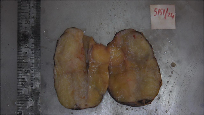
Case presentation: A male patient in his early 60s with a history of hypertension presented with an adrenal incidentaloma. Imaging revealed a well-defined, hypodense right adrenal mass (35 HU) with calcifications and an enlarged aortocaval lymph node, raising suspicion for carcinoma. Biochemical analysis showed a non-functional adrenal tumour. Intraoperatively, the mass appeared benign, but the lymph node deposit suggested malignancy. Histopathological examination confirmed right adrenal ganglioneuroma with metastatic deposits in the aortocaval lymph nodes. The patient recovered well postoperatively, with normal serum cortisol levels and no signs of recurrence at 6-month follow-up.
Conclusion: Adrenal ganglioneuromas can present as adrenal incidentalomas with lymph node involvement, mimicking malignancy. Surgery remains the gold standard for treatment, and postoperative histopathology is crucial for diagnosis. In such cases, no adjuvant therapy or stringent follow-up is required after surgical removal.

 求助内容:
求助内容: 应助结果提醒方式:


