Muhammad Mudasir Saleem, Mishal Pervaiz, Ismail Mazhar, Uswah Shoaib, Muhammad Umar Rafique
{"title":"Neonatal Adrenal Haemorrhage Mimicking Acute Scrotum: A Case Report.","authors":"Muhammad Mudasir Saleem, Mishal Pervaiz, Ismail Mazhar, Uswah Shoaib, Muhammad Umar Rafique","doi":"10.15190/d.2025.9","DOIUrl":null,"url":null,"abstract":"<p><p>Acute scrotum in neonates is a rare condition with multiple causes, including incarcerated hernia, testicular torsion, birth trauma, gross hydrocele, and neonatal adrenal haemorrhage, the least common aetiology. Early diagnosis and intervention are essential to prevent testicular ischemia. Due to the continuity between the retroperitoneum and the scrotum via the processus vaginalis and inguinal canal, blood from an adrenal haemorrhage may track down into the scrotum, leading to swelling and discoloration. We report a case of a 1-day-old male neonate born via emergency caesarean section at 37 weeks due to foetal distress. The baby initially admitted to the NICU for transient tachypnoea, developed a right hemi-scrotal swelling with bluish discoloration on the second day of life. Scrotal ultrasound suggested testicular torsion, but Doppler imaging showed absent blood flow. Further abdominal ultrasound confirmed a right adrenal haemorrhage. The neonate was managed conservatively with intravenous fluids, antibiotics, oxygen support, and coagulation management. Serial ultrasounds showed gradual resolution, and he was discharged on the 17th postnatal day. Follow-up at 1 and 3 months showed complete recovery with normal growth. Neonatal adrenal haemorrhage should be considered in cases of acute scrotum, especially in neonates with birth asphyxia. Abdominal ultrasound can aid in diagnosis, preventing diagnostic delays, unnecessary surgery, and anaesthesia exposure. This case highlights the importance of thorough imaging and awareness of rare differential diagnoses, contributing to improved clinical practice and better neonatal outcomes.</p>","PeriodicalId":72829,"journal":{"name":"Discoveries (Craiova, Romania)","volume":"13 1","pages":"e210"},"PeriodicalIF":0.0000,"publicationDate":"2025-06-30","publicationTypes":"Journal Article","fieldsOfStudy":null,"isOpenAccess":false,"openAccessPdf":"https://www.ncbi.nlm.nih.gov/pmc/articles/PMC12323913/pdf/","citationCount":"0","resultStr":null,"platform":"Semanticscholar","paperid":null,"PeriodicalName":"Discoveries (Craiova, Romania)","FirstCategoryId":"1085","ListUrlMain":"https://doi.org/10.15190/d.2025.9","RegionNum":0,"RegionCategory":null,"ArticlePicture":[],"TitleCN":null,"AbstractTextCN":null,"PMCID":null,"EPubDate":"2025/4/1 0:00:00","PubModel":"eCollection","JCR":"","JCRName":"","Score":null,"Total":0}
引用次数: 0
Abstract
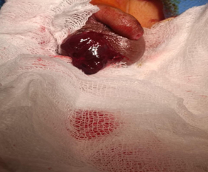
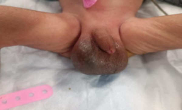
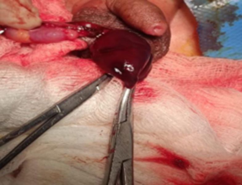
Acute scrotum in neonates is a rare condition with multiple causes, including incarcerated hernia, testicular torsion, birth trauma, gross hydrocele, and neonatal adrenal haemorrhage, the least common aetiology. Early diagnosis and intervention are essential to prevent testicular ischemia. Due to the continuity between the retroperitoneum and the scrotum via the processus vaginalis and inguinal canal, blood from an adrenal haemorrhage may track down into the scrotum, leading to swelling and discoloration. We report a case of a 1-day-old male neonate born via emergency caesarean section at 37 weeks due to foetal distress. The baby initially admitted to the NICU for transient tachypnoea, developed a right hemi-scrotal swelling with bluish discoloration on the second day of life. Scrotal ultrasound suggested testicular torsion, but Doppler imaging showed absent blood flow. Further abdominal ultrasound confirmed a right adrenal haemorrhage. The neonate was managed conservatively with intravenous fluids, antibiotics, oxygen support, and coagulation management. Serial ultrasounds showed gradual resolution, and he was discharged on the 17th postnatal day. Follow-up at 1 and 3 months showed complete recovery with normal growth. Neonatal adrenal haemorrhage should be considered in cases of acute scrotum, especially in neonates with birth asphyxia. Abdominal ultrasound can aid in diagnosis, preventing diagnostic delays, unnecessary surgery, and anaesthesia exposure. This case highlights the importance of thorough imaging and awareness of rare differential diagnoses, contributing to improved clinical practice and better neonatal outcomes.

 求助内容:
求助内容: 应助结果提醒方式:


