{"title":"Deep-learning reconstruction of the prostate improves image quality and acquisition time in T2-weighted imaging.","authors":"Daichi Kobayashi, Hayato Tomita, Tsuyoshi Morimoto, Yuki Deguchi, Hirofumi Fukuchi, Hikaru Ishida, Kumie Miyakawa, Yasuyuki Kobayashi, Hidefumi Mimura","doi":"10.18999/nagjms.87.2.264","DOIUrl":null,"url":null,"abstract":"<p><p>We compared the qualitative and quantitative quality of prostate conventional T2-weighted imaging and T2-weighted imaging with deep-learning reconstruction. Patients with suspected prostate cancer undergoing magnetic resonance imaging between April 2022 and June 2023 were included. Quantitative analysis was performed to determine the signal-to-noise and contrast ratios of the perirectal fat tissue, internal obturator muscle, and pubic tubercle. Eight periprostatic anatomical structures, overall image quality, and motion artifacts were evaluated by two radiologists using 5- or 4-point scales. Qualitative analysis results were compared to determine the agreement between the two radiologists. In total, 106 patients (mean age: 71 ± 8.3 years; 106 men) were included in this study. The acquisition time for conventional T2-weighted imaging and T2-weighted imaging with deep-learning reconstruction was 4 min and 16 s and 2 min and 12 s, respectively. The signal-to-noise ratio of the perirectal fat tissue and internal obturator muscle and contrast ratio of fat/muscle and bone/muscle determined via T2-weighted imaging with deep-learning reconstruction were significantly superior to those determined via conventional T2-weighted imaging (both <i>p</i> < 0.01). Compared with conventional T2-weighted imaging, T2-weighted imaging with deep-learning reconstruction showed significant improvement in the visualization of the periprostatic anatomy, overall image quality, and motion artifacts (both <i>p</i> < 0.05). Compared with conventional methods, T2-weighted imaging with deep-learning reconstruction facilitated the acquisition of good-quality magnetic resonance images of the prostate within a shorter acquisition time. T2-weighted imaging with deep-learning reconstruction will aid clinicians in diagnosing prostate cancer with shortened acquisition time while maintaining quantitative and qualitative image properties.</p>","PeriodicalId":49014,"journal":{"name":"Nagoya Journal of Medical Science","volume":"87 2","pages":"264-271"},"PeriodicalIF":0.9000,"publicationDate":"2025-05-01","publicationTypes":"Journal Article","fieldsOfStudy":null,"isOpenAccess":false,"openAccessPdf":"https://www.ncbi.nlm.nih.gov/pmc/articles/PMC12320318/pdf/","citationCount":"0","resultStr":null,"platform":"Semanticscholar","paperid":null,"PeriodicalName":"Nagoya Journal of Medical Science","FirstCategoryId":"3","ListUrlMain":"https://doi.org/10.18999/nagjms.87.2.264","RegionNum":4,"RegionCategory":"医学","ArticlePicture":[],"TitleCN":null,"AbstractTextCN":null,"PMCID":null,"EPubDate":"","PubModel":"","JCR":"Q4","JCRName":"MEDICINE, RESEARCH & EXPERIMENTAL","Score":null,"Total":0}
引用次数: 0
Abstract
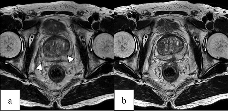
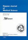
We compared the qualitative and quantitative quality of prostate conventional T2-weighted imaging and T2-weighted imaging with deep-learning reconstruction. Patients with suspected prostate cancer undergoing magnetic resonance imaging between April 2022 and June 2023 were included. Quantitative analysis was performed to determine the signal-to-noise and contrast ratios of the perirectal fat tissue, internal obturator muscle, and pubic tubercle. Eight periprostatic anatomical structures, overall image quality, and motion artifacts were evaluated by two radiologists using 5- or 4-point scales. Qualitative analysis results were compared to determine the agreement between the two radiologists. In total, 106 patients (mean age: 71 ± 8.3 years; 106 men) were included in this study. The acquisition time for conventional T2-weighted imaging and T2-weighted imaging with deep-learning reconstruction was 4 min and 16 s and 2 min and 12 s, respectively. The signal-to-noise ratio of the perirectal fat tissue and internal obturator muscle and contrast ratio of fat/muscle and bone/muscle determined via T2-weighted imaging with deep-learning reconstruction were significantly superior to those determined via conventional T2-weighted imaging (both p < 0.01). Compared with conventional T2-weighted imaging, T2-weighted imaging with deep-learning reconstruction showed significant improvement in the visualization of the periprostatic anatomy, overall image quality, and motion artifacts (both p < 0.05). Compared with conventional methods, T2-weighted imaging with deep-learning reconstruction facilitated the acquisition of good-quality magnetic resonance images of the prostate within a shorter acquisition time. T2-weighted imaging with deep-learning reconstruction will aid clinicians in diagnosing prostate cancer with shortened acquisition time while maintaining quantitative and qualitative image properties.
期刊介绍:
The Journal publishes original papers in the areas of medical science and its related fields. Reviews, symposium reports, short communications, notes, case reports, hypothesis papers, medical image at a glance, video and announcements are also accepted.
Manuscripts should be in English. It is recommended that an English check of the manuscript by a competent and knowledgeable native speaker be completed before submission.

 求助内容:
求助内容: 应助结果提醒方式:


