{"title":"Glymphatic dysfunction associated with cortisol dysregulation in major depressive disorder.","authors":"Shengli Chen, Ziyun Xu, Zheng Guo, Shiwei Lin, Hongyuan Zhang, Qunjun Liang, Qianyun Chen, Yingli Zhang, Bo Peng, Yanzhen Zhao, Gangqiang Hou, Yingwei Qiu","doi":"10.1038/s41398-025-03486-1","DOIUrl":null,"url":null,"abstract":"<p><p>Cortisol dysregulation plays a critical role in the pathophysiology of depression, but its exact impact on the brain remains unclear. In this cross-sectional study, 210 participants, including 164 depressed patients and 46 healthy controls (HCs), were assessed. Glymphatic circulation was evaluated on Magnetic Resonance Imaging via choroid plexus (CP) volume fraction, perivascular space (PVS) volume fraction, fractional volume of extracellular-free water (FW), and diffusion tensor imaging along the perivascular space (DTI-ALPS) index. Chemiluminescence was employed to analyze the cortisol levels in a sub-cohort of the patients. Independent sample t-tests and Pearson's correlation analysis were used to assess differences in these metrics between groups and their correlation with cortisol levels. After adjusting for age, sex, years of education, and total intracranial volume, depressed patients exhibited a significantly higher FW and lower ALPS than HCs. No significant differences were found in CP volume and PVS fraction between depressed patients and HCs. Additionally, the increased FW correlated positively with cortisol levels in depressed patients, suggesting that glymphatic dysfunction is linked to plasma cortisol levels in depression.</p>","PeriodicalId":23278,"journal":{"name":"Translational Psychiatry","volume":"15 1","pages":"265"},"PeriodicalIF":6.2000,"publicationDate":"2025-08-05","publicationTypes":"Journal Article","fieldsOfStudy":null,"isOpenAccess":false,"openAccessPdf":"https://www.ncbi.nlm.nih.gov/pmc/articles/PMC12325902/pdf/","citationCount":"0","resultStr":null,"platform":"Semanticscholar","paperid":null,"PeriodicalName":"Translational Psychiatry","FirstCategoryId":"3","ListUrlMain":"https://doi.org/10.1038/s41398-025-03486-1","RegionNum":1,"RegionCategory":"医学","ArticlePicture":[],"TitleCN":null,"AbstractTextCN":null,"PMCID":null,"EPubDate":"","PubModel":"","JCR":"Q1","JCRName":"PSYCHIATRY","Score":null,"Total":0}
引用次数: 0
Abstract
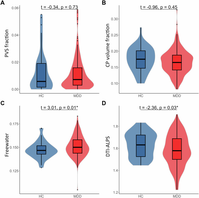
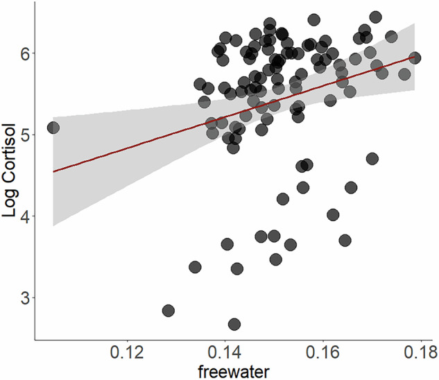
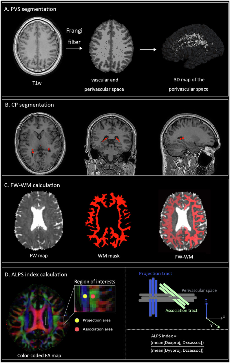
Cortisol dysregulation plays a critical role in the pathophysiology of depression, but its exact impact on the brain remains unclear. In this cross-sectional study, 210 participants, including 164 depressed patients and 46 healthy controls (HCs), were assessed. Glymphatic circulation was evaluated on Magnetic Resonance Imaging via choroid plexus (CP) volume fraction, perivascular space (PVS) volume fraction, fractional volume of extracellular-free water (FW), and diffusion tensor imaging along the perivascular space (DTI-ALPS) index. Chemiluminescence was employed to analyze the cortisol levels in a sub-cohort of the patients. Independent sample t-tests and Pearson's correlation analysis were used to assess differences in these metrics between groups and their correlation with cortisol levels. After adjusting for age, sex, years of education, and total intracranial volume, depressed patients exhibited a significantly higher FW and lower ALPS than HCs. No significant differences were found in CP volume and PVS fraction between depressed patients and HCs. Additionally, the increased FW correlated positively with cortisol levels in depressed patients, suggesting that glymphatic dysfunction is linked to plasma cortisol levels in depression.
期刊介绍:
Psychiatry has suffered tremendously by the limited translational pipeline. Nobel laureate Julius Axelrod''s discovery in 1961 of monoamine reuptake by pre-synaptic neurons still forms the basis of contemporary antidepressant treatment. There is a grievous gap between the explosion of knowledge in neuroscience and conceptually novel treatments for our patients. Translational Psychiatry bridges this gap by fostering and highlighting the pathway from discovery to clinical applications, healthcare and global health. We view translation broadly as the full spectrum of work that marks the pathway from discovery to global health, inclusive. The steps of translation that are within the scope of Translational Psychiatry include (i) fundamental discovery, (ii) bench to bedside, (iii) bedside to clinical applications (clinical trials), (iv) translation to policy and health care guidelines, (v) assessment of health policy and usage, and (vi) global health. All areas of medical research, including — but not restricted to — molecular biology, genetics, pharmacology, imaging and epidemiology are welcome as they contribute to enhance the field of translational psychiatry.


 求助内容:
求助内容: 应助结果提醒方式:


