Eun Ji Han, Ji Won Moon, Ji Min Son, Mihai Oltean, Mats Hellström, Francesco Boccardo, Min Jong Song
{"title":"Predicting the Outcome of Lymphovenous Anastomosis for Lower Extremity Lymphedema through Lymphoscintigraphy.","authors":"Eun Ji Han, Ji Won Moon, Ji Min Son, Mihai Oltean, Mats Hellström, Francesco Boccardo, Min Jong Song","doi":"10.7150/ijms.111506","DOIUrl":null,"url":null,"abstract":"<p><p><b>Background:</b> Lymphovenous anastomosis (LVA) is an effective treatment for restoring lymphatic function in patients with lymphedema. This study aimed to assess the predictive value of preoperative lymphoscintigraphy in female patients undergoing LVA for lower extremity lymphedema (LEL). <b>Methods:</b> Female patients with unilateral LEL who underwent preoperative lymphoscintigraphy followed by LVA were retrospectively examined. In the lymphoscintigraphy, the transport index (TI) was calculated based on five visual interpretation criteria: lymphatic transport kinetics, dermal backflow pattern, time to appearance of lymph nodes, visualization of lymph nodes, and visualization of vessels. For volume assessment, the LEL index (LELI) was calculated as the sum of circumferences at five predefined sites of the lower extremity, divided by body mass index. LELI was measured before and after LVA at 1, 3, and 6 months. Postoperative changes in LELI were compared with preoperative variables, including TI. <b>Results:</b> The study included 45 female patients (mean age 56 ± 10 years) with unilateral LEL, of whom 78% had clinical stage 3 lymphedema. The mean TI of the affected lower extremities at 240 and 120 min was 25.5 ± 11.0 and 26.5 ± 11.1, respectively. TI was significantly associated with clinical stage and preoperative volume excess. Postoperatively, the mean LELI reduction was 7 ± 5% at 1 month, 8 ± 5% at 3 months, and 6 ± 7% at 6 months. Significant negative correlations were found between the TI at both 240 and 120 min and postoperative LELI changes at 3 and 6 months (p < 0.05). <b>Conclusions:</b> Preoperative lymphoscintigraphy, specifically the TI, is valuable for assessing the severity of lymphedema and predicting short-term outcomes of LVA in female patients with LEL. The TI can be calculated from lymphoscintigraphy performed up to 2 h.</p>","PeriodicalId":14031,"journal":{"name":"International Journal of Medical Sciences","volume":"22 13","pages":"3174-3181"},"PeriodicalIF":3.2000,"publicationDate":"2025-06-23","publicationTypes":"Journal Article","fieldsOfStudy":null,"isOpenAccess":false,"openAccessPdf":"https://www.ncbi.nlm.nih.gov/pmc/articles/PMC12320694/pdf/","citationCount":"0","resultStr":null,"platform":"Semanticscholar","paperid":null,"PeriodicalName":"International Journal of Medical Sciences","FirstCategoryId":"3","ListUrlMain":"https://doi.org/10.7150/ijms.111506","RegionNum":3,"RegionCategory":"医学","ArticlePicture":[],"TitleCN":null,"AbstractTextCN":null,"PMCID":null,"EPubDate":"2025/1/1 0:00:00","PubModel":"eCollection","JCR":"Q1","JCRName":"MEDICINE, GENERAL & INTERNAL","Score":null,"Total":0}
引用次数: 0
Abstract
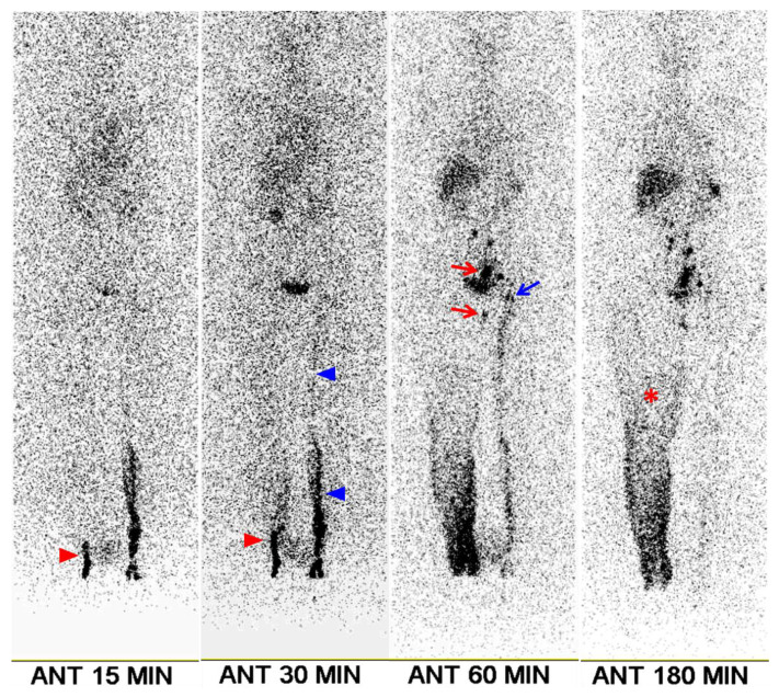
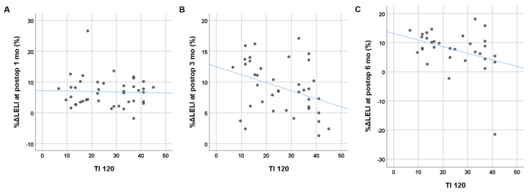
Background: Lymphovenous anastomosis (LVA) is an effective treatment for restoring lymphatic function in patients with lymphedema. This study aimed to assess the predictive value of preoperative lymphoscintigraphy in female patients undergoing LVA for lower extremity lymphedema (LEL). Methods: Female patients with unilateral LEL who underwent preoperative lymphoscintigraphy followed by LVA were retrospectively examined. In the lymphoscintigraphy, the transport index (TI) was calculated based on five visual interpretation criteria: lymphatic transport kinetics, dermal backflow pattern, time to appearance of lymph nodes, visualization of lymph nodes, and visualization of vessels. For volume assessment, the LEL index (LELI) was calculated as the sum of circumferences at five predefined sites of the lower extremity, divided by body mass index. LELI was measured before and after LVA at 1, 3, and 6 months. Postoperative changes in LELI were compared with preoperative variables, including TI. Results: The study included 45 female patients (mean age 56 ± 10 years) with unilateral LEL, of whom 78% had clinical stage 3 lymphedema. The mean TI of the affected lower extremities at 240 and 120 min was 25.5 ± 11.0 and 26.5 ± 11.1, respectively. TI was significantly associated with clinical stage and preoperative volume excess. Postoperatively, the mean LELI reduction was 7 ± 5% at 1 month, 8 ± 5% at 3 months, and 6 ± 7% at 6 months. Significant negative correlations were found between the TI at both 240 and 120 min and postoperative LELI changes at 3 and 6 months (p < 0.05). Conclusions: Preoperative lymphoscintigraphy, specifically the TI, is valuable for assessing the severity of lymphedema and predicting short-term outcomes of LVA in female patients with LEL. The TI can be calculated from lymphoscintigraphy performed up to 2 h.
期刊介绍:
Original research papers, reviews, and short research communications in any medical related area can be submitted to the Journal on the understanding that the work has not been published previously in whole or part and is not under consideration for publication elsewhere. Manuscripts in basic science and clinical medicine are both considered. There is no restriction on the length of research papers and reviews, although authors are encouraged to be concise. Short research communication is limited to be under 2500 words.
 求助内容:
求助内容: 应助结果提醒方式:


