{"title":"Preoperative MRI and CA19-9 for predicting occult lymph node metastasis in small pancreatic ductal adenocarcinoma (≤ 2 cm).","authors":"Qiying Tang, Lei Li, Zhiwei Pan, Jianbo Li, Xiaolan Huang, Mengsu Zeng, Haitao Sun, Jianjun Zhou","doi":"10.1186/s12880-025-01854-3","DOIUrl":null,"url":null,"abstract":"<p><strong>Aim: </strong>Accurate prediction of occult lymph node metastasis (OLNM) in small pancreatic ductal adenocarcinoma (sPDAC) (≤ 2 cm) is crucial for curative management. This study aims to explore clinical and MRI features associated with OLNM in sPDAC and their pathological and prognostic implications.</p><p><strong>Materials and methods: </strong>This retrospective study included 135 patients with pathologically confirmed sPDAC who underwent surgery between September 2014 and September 2023. Preoperative multi-sequence MRI, clinical data, and pathological features were analyzed. Univariate and multivariate logistic regression models were used to identify risk predictors of OLNM in sPDAC. Receiver operating characteristic (ROC) analysis was performed to assess diagnostic performance and Kaplan-Meier survival analysis was used to evaluate prognostic outcomes.</p><p><strong>Results: </strong>OLNM was present in 43 (31.9%) sPDAC patients. Univariate and multivariate analysis identified elevated CA19-9 (> 100 U/mL) (OR = 2.404, P = 0.040) and low apparent diffusion coefficient (ADC) values (OR = 0.243, P = 0.031) as independent predictors of OLNM. The combined clinical-radiological model demonstrated an AUC of 0.740, significantly higher than CA19-9 (AUC = 0.653, P = 0.021) or ADC alone (AUC = 0.635, P = 0.035). sPDAC patients with OLNM exhibited higher rates of lymphovascular invasion (44.2%, P = 0.013) and pathological fat invasion (86.0%, P = 0.030). OLNM was associated with significantly worse OS and DFS (P = 0.034 and 0.043).</p><p><strong>Conclusions: </strong>OLNM is associated with adverse pathological features and poorer prognosis. The combination of preoperative MRI assessment of ADC and CA19-9 may aid in identifying sPDAC patients at high risk for OLNM.</p><p><strong>Clinical trial number: </strong>Not applicable.</p>","PeriodicalId":9020,"journal":{"name":"BMC Medical Imaging","volume":"25 1","pages":"318"},"PeriodicalIF":3.2000,"publicationDate":"2025-08-06","publicationTypes":"Journal Article","fieldsOfStudy":null,"isOpenAccess":false,"openAccessPdf":"https://www.ncbi.nlm.nih.gov/pmc/articles/PMC12326715/pdf/","citationCount":"0","resultStr":null,"platform":"Semanticscholar","paperid":null,"PeriodicalName":"BMC Medical Imaging","FirstCategoryId":"3","ListUrlMain":"https://doi.org/10.1186/s12880-025-01854-3","RegionNum":3,"RegionCategory":"医学","ArticlePicture":[],"TitleCN":null,"AbstractTextCN":null,"PMCID":null,"EPubDate":"","PubModel":"","JCR":"Q2","JCRName":"RADIOLOGY, NUCLEAR MEDICINE & MEDICAL IMAGING","Score":null,"Total":0}
引用次数: 0
Abstract
Aim: Accurate prediction of occult lymph node metastasis (OLNM) in small pancreatic ductal adenocarcinoma (sPDAC) (≤ 2 cm) is crucial for curative management. This study aims to explore clinical and MRI features associated with OLNM in sPDAC and their pathological and prognostic implications.
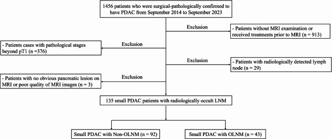
Materials and methods: This retrospective study included 135 patients with pathologically confirmed sPDAC who underwent surgery between September 2014 and September 2023. Preoperative multi-sequence MRI, clinical data, and pathological features were analyzed. Univariate and multivariate logistic regression models were used to identify risk predictors of OLNM in sPDAC. Receiver operating characteristic (ROC) analysis was performed to assess diagnostic performance and Kaplan-Meier survival analysis was used to evaluate prognostic outcomes.
Results: OLNM was present in 43 (31.9%) sPDAC patients. Univariate and multivariate analysis identified elevated CA19-9 (> 100 U/mL) (OR = 2.404, P = 0.040) and low apparent diffusion coefficient (ADC) values (OR = 0.243, P = 0.031) as independent predictors of OLNM. The combined clinical-radiological model demonstrated an AUC of 0.740, significantly higher than CA19-9 (AUC = 0.653, P = 0.021) or ADC alone (AUC = 0.635, P = 0.035). sPDAC patients with OLNM exhibited higher rates of lymphovascular invasion (44.2%, P = 0.013) and pathological fat invasion (86.0%, P = 0.030). OLNM was associated with significantly worse OS and DFS (P = 0.034 and 0.043).
Conclusions: OLNM is associated with adverse pathological features and poorer prognosis. The combination of preoperative MRI assessment of ADC and CA19-9 may aid in identifying sPDAC patients at high risk for OLNM.
期刊介绍:
BMC Medical Imaging is an open access journal publishing original peer-reviewed research articles in the development, evaluation, and use of imaging techniques and image processing tools to diagnose and manage disease.


 求助内容:
求助内容: 应助结果提醒方式:


