Céline van Haaren, Bernadette Byrne and Sergei G. Kazarian
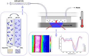
{"title":"Assessment of IgG stability in a low pH elution buffer using ATR-FTIR spectroscopic imaging and microfluidics","authors":"Céline van Haaren, Bernadette Byrne and Sergei G. Kazarian","doi":"10.1039/D5AN00664C","DOIUrl":null,"url":null,"abstract":"<p >Monoclonal antibodies (mAbs) represent the largest class of biopharmaceuticals, playing a vital role in the treatment of a wide range of diseases. Although the production of high quality mAbs has significantly improved over the last three decades, particularly in terms of scale and yield, the antibody's complex nature poses several challenges during bioprocessing. One of the main challenges in the production of mAbs is the formation of aggregates, which may cause harmful immunogenic responses in patients if not removed from the final drug product. Exposure to a low pH environment during protein A chromatography and viral inactivation is thought to be the major contributor to aggregate formation and has therefore been a topic of study for many years. Here, we investigate the stability of an IgG4 mAb in a low pH elution buffer (pH 3.5) under flow using ATR-FTIR spectroscopic imaging. This method, making use of a microfluidic set-up, enables non-destructive monitoring of mAb structural stability under bioprocessing-relevant conditions. Samples were (i) prepared through dialysis into the elution buffer and (ii) collected directly after elution from the protein A column, after which their stability was assessed under flow at two different temperatures (30 °C and 45 °C). Spectroscopic images and associated IR absorption spectra revealed that in both cases the protein in the low pH buffer underwent small, but measurable, structural changes at 30 °C. However, at 45 °C, the protein rapidly aggregated as indicated by a major shift in the Amide I peak position from 1637 cm<small><sup>−1</sup></small> to 1625 cm<small><sup>−1</sup></small>, representing formation of inter-molecular beta sheets. These results confirm the destabilising effect of the low pH environment and demonstrate the applicability of ATR-FTIR spectroscopic imaging in combination with microfluidics as a powerful analytical tool for the analysis of protein structural stability under flow.</p>","PeriodicalId":63,"journal":{"name":"Analyst","volume":" 18","pages":" 4201-4210"},"PeriodicalIF":3.3000,"publicationDate":"2025-08-05","publicationTypes":"Journal Article","fieldsOfStudy":null,"isOpenAccess":false,"openAccessPdf":"https://pubs.rsc.org/en/content/articlepdf/2025/an/d5an00664c?page=search","citationCount":"0","resultStr":null,"platform":"Semanticscholar","paperid":null,"PeriodicalName":"Analyst","FirstCategoryId":"92","ListUrlMain":"https://pubs.rsc.org/en/content/articlelanding/2025/an/d5an00664c","RegionNum":3,"RegionCategory":"化学","ArticlePicture":[],"TitleCN":null,"AbstractTextCN":null,"PMCID":null,"EPubDate":"","PubModel":"","JCR":"Q2","JCRName":"CHEMISTRY, ANALYTICAL","Score":null,"Total":0}
引用次数: 0
Abstract
Monoclonal antibodies (mAbs) represent the largest class of biopharmaceuticals, playing a vital role in the treatment of a wide range of diseases. Although the production of high quality mAbs has significantly improved over the last three decades, particularly in terms of scale and yield, the antibody's complex nature poses several challenges during bioprocessing. One of the main challenges in the production of mAbs is the formation of aggregates, which may cause harmful immunogenic responses in patients if not removed from the final drug product. Exposure to a low pH environment during protein A chromatography and viral inactivation is thought to be the major contributor to aggregate formation and has therefore been a topic of study for many years. Here, we investigate the stability of an IgG4 mAb in a low pH elution buffer (pH 3.5) under flow using ATR-FTIR spectroscopic imaging. This method, making use of a microfluidic set-up, enables non-destructive monitoring of mAb structural stability under bioprocessing-relevant conditions. Samples were (i) prepared through dialysis into the elution buffer and (ii) collected directly after elution from the protein A column, after which their stability was assessed under flow at two different temperatures (30 °C and 45 °C). Spectroscopic images and associated IR absorption spectra revealed that in both cases the protein in the low pH buffer underwent small, but measurable, structural changes at 30 °C. However, at 45 °C, the protein rapidly aggregated as indicated by a major shift in the Amide I peak position from 1637 cm−1 to 1625 cm−1, representing formation of inter-molecular beta sheets. These results confirm the destabilising effect of the low pH environment and demonstrate the applicability of ATR-FTIR spectroscopic imaging in combination with microfluidics as a powerful analytical tool for the analysis of protein structural stability under flow.
 求助内容:
求助内容: 应助结果提醒方式:


