Hailey Mattheisen, Abigail Peterson, Brandon Laing, Nathan T Zwagerman, Michael S Harris, Stephanie Cheok
{"title":"Life-threatening pneumocephalus from middle ear defect causing cerebrospinal fluid leakage into the eustachian tube: illustrative case.","authors":"Hailey Mattheisen, Abigail Peterson, Brandon Laing, Nathan T Zwagerman, Michael S Harris, Stephanie Cheok","doi":"10.3171/CASE24834","DOIUrl":null,"url":null,"abstract":"<p><strong>Background: </strong>Spontaneous pneumocephalus is a rare, but potentially serious condition often secondary to a CSF leak. This egress of CSF causes an inward movement of air to replace the lost volume. CSF leaks are typically posttraumatic and present as rhinorrhea or otorrhea. Locating the bony defect and herniating contents through imaging is crucial for planned surgical intervention. In this report, the author present the case of a patient with progressive spontaneous pneumocephalus with an apparent but unidentifiable CSF leak.</p><p><strong>Observations: </strong>A 76-year-old female presented to the authors' institution with rapidly progressing spontaneous pneumocephalus. This case is unique in that the patient's pneumocephalus continued to expand without any radiological indication of extracranial CSF, leading us to believe that the CSF was intermittently leaking through the eustachian tube and passing down the patient's oropharynx. Given the presumed pathway, an initial conservative surgical approach was chosen for this patient's spontaneous pneumocephalus instead of the traditional middle cranial fossa or transmastoid approach.</p><p><strong>Lessons: </strong>To repair the leak, the authors attempted a more conservative approach involving closure of the external acoustic meatus and obliteration of the eustachian tube. A few studies using a similar procedure reported a lower risk of complications and reduction in postoperative CSF leaks. https://thejns.org/doi/10.3171/CASE24834.</p>","PeriodicalId":94098,"journal":{"name":"Journal of neurosurgery. Case lessons","volume":"10 5","pages":""},"PeriodicalIF":0.0000,"publicationDate":"2025-08-04","publicationTypes":"Journal Article","fieldsOfStudy":null,"isOpenAccess":false,"openAccessPdf":"https://www.ncbi.nlm.nih.gov/pmc/articles/PMC12320727/pdf/","citationCount":"0","resultStr":null,"platform":"Semanticscholar","paperid":null,"PeriodicalName":"Journal of neurosurgery. Case lessons","FirstCategoryId":"1085","ListUrlMain":"https://doi.org/10.3171/CASE24834","RegionNum":0,"RegionCategory":null,"ArticlePicture":[],"TitleCN":null,"AbstractTextCN":null,"PMCID":null,"EPubDate":"","PubModel":"","JCR":"","JCRName":"","Score":null,"Total":0}
引用次数: 0
Abstract
Background: Spontaneous pneumocephalus is a rare, but potentially serious condition often secondary to a CSF leak. This egress of CSF causes an inward movement of air to replace the lost volume. CSF leaks are typically posttraumatic and present as rhinorrhea or otorrhea. Locating the bony defect and herniating contents through imaging is crucial for planned surgical intervention. In this report, the author present the case of a patient with progressive spontaneous pneumocephalus with an apparent but unidentifiable CSF leak.
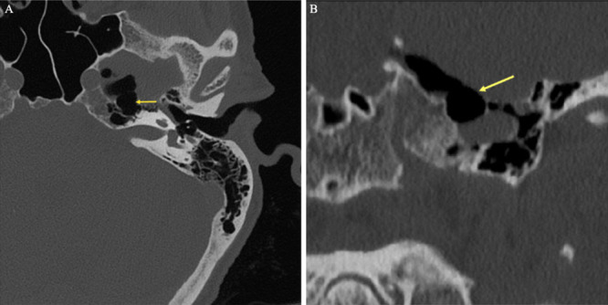
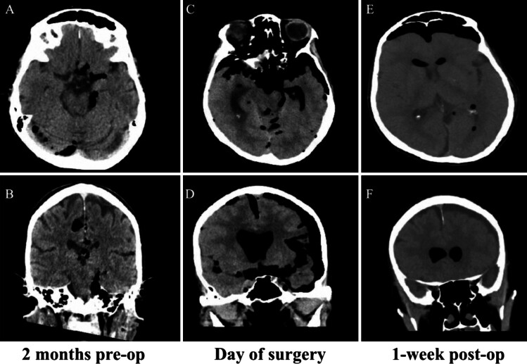
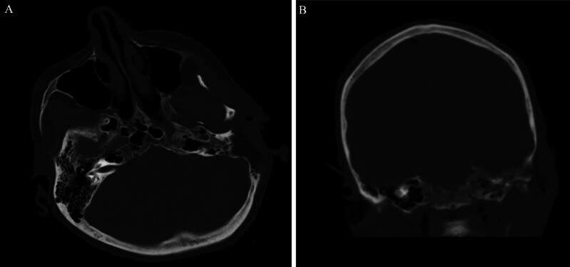
Observations: A 76-year-old female presented to the authors' institution with rapidly progressing spontaneous pneumocephalus. This case is unique in that the patient's pneumocephalus continued to expand without any radiological indication of extracranial CSF, leading us to believe that the CSF was intermittently leaking through the eustachian tube and passing down the patient's oropharynx. Given the presumed pathway, an initial conservative surgical approach was chosen for this patient's spontaneous pneumocephalus instead of the traditional middle cranial fossa or transmastoid approach.
Lessons: To repair the leak, the authors attempted a more conservative approach involving closure of the external acoustic meatus and obliteration of the eustachian tube. A few studies using a similar procedure reported a lower risk of complications and reduction in postoperative CSF leaks. https://thejns.org/doi/10.3171/CASE24834.

 求助内容:
求助内容: 应助结果提醒方式:


