Lei Xiang, Rouyan Chen, Joanne Tsui Ming Tan, Victoria Nankivell, Christina A Bursill, Robert A McLaughlin, Jiawen Li
{"title":"Identification and removal of system-induced autofluorescence in miniaturized fiber-optic fluorescence endoscopes.","authors":"Lei Xiang, Rouyan Chen, Joanne Tsui Ming Tan, Victoria Nankivell, Christina A Bursill, Robert A McLaughlin, Jiawen Li","doi":"10.1093/pnasnexus/pgaf226","DOIUrl":null,"url":null,"abstract":"<p><p>Miniaturized fiber-optic fluorescence endoscopes play a crucial role in medical diagnostics and research, but system-induced autofluorescence remains a significant challenge, particularly in single-fiber setups. While recent advances, such as double-clad fiber (DCF) and DCF couplers, have reduced background noise, complete elimination remains challenging. Research on the various sources of system-induced autofluorescence and the methods to remove them is scarce. This study seeks to fulfill this need by proposing practical approaches to the removal of system-induced autofluorescence. This study presents the methods to suppress static background noise and proposes an algorithm based on least-squares linear spectral unmixing to remove variable system-induced autofluorescence artifacts. The algorithm was evaluated on a single-fiber DCF intravascular imaging system, with phantom and rodent in vivo experiments confirming its effectiveness. Results showed accurate differentiation between true sample fluorescence and system-induced autofluorescence artifacts through the validation with optical coherence tomography images and histology results, further verified by statistical analysis. Unlike simple background subtraction, the method addresses both background noise and incidental artifacts, providing robust performance under varying conditions. Our method may be adapted to various fiber-based endoscopy setups and be compatible with different fluorescent agents and autofluorescence imaging, broadening its applicability in biomedical imaging.</p>","PeriodicalId":74468,"journal":{"name":"PNAS nexus","volume":"4 8","pages":"pgaf226"},"PeriodicalIF":3.8000,"publicationDate":"2025-07-23","publicationTypes":"Journal Article","fieldsOfStudy":null,"isOpenAccess":false,"openAccessPdf":"https://www.ncbi.nlm.nih.gov/pmc/articles/PMC12318715/pdf/","citationCount":"0","resultStr":null,"platform":"Semanticscholar","paperid":null,"PeriodicalName":"PNAS nexus","FirstCategoryId":"1085","ListUrlMain":"https://doi.org/10.1093/pnasnexus/pgaf226","RegionNum":0,"RegionCategory":null,"ArticlePicture":[],"TitleCN":null,"AbstractTextCN":null,"PMCID":null,"EPubDate":"2025/8/1 0:00:00","PubModel":"eCollection","JCR":"Q2","JCRName":"MULTIDISCIPLINARY SCIENCES","Score":null,"Total":0}
引用次数: 0
Abstract
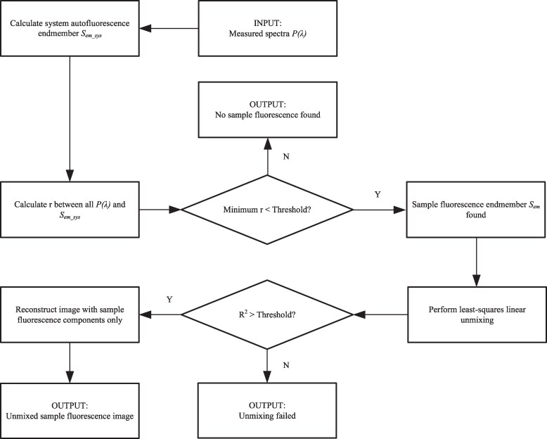
Miniaturized fiber-optic fluorescence endoscopes play a crucial role in medical diagnostics and research, but system-induced autofluorescence remains a significant challenge, particularly in single-fiber setups. While recent advances, such as double-clad fiber (DCF) and DCF couplers, have reduced background noise, complete elimination remains challenging. Research on the various sources of system-induced autofluorescence and the methods to remove them is scarce. This study seeks to fulfill this need by proposing practical approaches to the removal of system-induced autofluorescence. This study presents the methods to suppress static background noise and proposes an algorithm based on least-squares linear spectral unmixing to remove variable system-induced autofluorescence artifacts. The algorithm was evaluated on a single-fiber DCF intravascular imaging system, with phantom and rodent in vivo experiments confirming its effectiveness. Results showed accurate differentiation between true sample fluorescence and system-induced autofluorescence artifacts through the validation with optical coherence tomography images and histology results, further verified by statistical analysis. Unlike simple background subtraction, the method addresses both background noise and incidental artifacts, providing robust performance under varying conditions. Our method may be adapted to various fiber-based endoscopy setups and be compatible with different fluorescent agents and autofluorescence imaging, broadening its applicability in biomedical imaging.

 求助内容:
求助内容: 应助结果提醒方式:


