Mohmed Hussien Ahmed Mohmed, Isra Hamed Dafallah Idrees, Ahmed Mostafa, Khalid Elfatih Mohammed Ibrahim, Amna Adnan Ahmad, Mahmoud H S Daoud
{"title":"A case report of long-segment tuberculous myelitis with concomitant tuberculous meningitis.","authors":"Mohmed Hussien Ahmed Mohmed, Isra Hamed Dafallah Idrees, Ahmed Mostafa, Khalid Elfatih Mohammed Ibrahim, Amna Adnan Ahmad, Mahmoud H S Daoud","doi":"10.21037/acr-24-211","DOIUrl":null,"url":null,"abstract":"<p><strong>Background: </strong>Tuberculous myelitis is a form of central nervous system tuberculosis (TB) that can be associated with intracranial involvement but rarely presents with extensive longitudinal involvement of more than one segment. We are reporting a case with tuberculous meningitis and long-segment myelitis in a previously undiagnosed patient with TB.</p><p><strong>Case description: </strong>A 53-year-old hypertensive male, presented with subacute lower limbs weakness, sensory level below his nipples, and urine retention. Erythrocyte sedimentation rate (ESR) was above 112 mm/hour. Magnetic resonance imaging (MRI) spine showed a long segment of hyperintense signal seen on the T2-weighted image (T2WI) images in the spinal cord extending from C6 to D3 vertebral segments, with heterogeneous post-contrast enhancement. Cerebrospinal fluid (CSF) analysis showed lymphocytic pleocytosis with high protein and low glucose, and polymerase chain reaction (PCR) for <i>Mycobacterium tuberculosis</i> (MBTB) was positive. The patient received intravenous methylprednisolone daily for 5 days and standard anti-TB medications [rifampicin, isoniazid (INH), pyrazinamide, and ethambutol] for 12 months. However, repeated CSF analysis 3 months after starting anti-TB medications showed a negative PCR for MBTB, normal cell count, and glucose with slightly elevated protein. Still, the patient did not show any clinical improvement.</p><p><strong>Conclusions: </strong>Long-segment tuberculous myelitis (LSTM) is a rare form of central nervous system TB that can be accompanied by tuberculous meningitis. It must be considered a differential diagnosis of neuromyelitis optica spectrum disorder (NMOSD), especially in endemic areas of TB, as the management approach is completely different.</p>","PeriodicalId":29752,"journal":{"name":"AME Case Reports","volume":"9 ","pages":"76"},"PeriodicalIF":0.7000,"publicationDate":"2025-06-06","publicationTypes":"Journal Article","fieldsOfStudy":null,"isOpenAccess":false,"openAccessPdf":"https://www.ncbi.nlm.nih.gov/pmc/articles/PMC12319586/pdf/","citationCount":"0","resultStr":null,"platform":"Semanticscholar","paperid":null,"PeriodicalName":"AME Case Reports","FirstCategoryId":"1085","ListUrlMain":"https://doi.org/10.21037/acr-24-211","RegionNum":0,"RegionCategory":null,"ArticlePicture":[],"TitleCN":null,"AbstractTextCN":null,"PMCID":null,"EPubDate":"2025/1/1 0:00:00","PubModel":"eCollection","JCR":"Q3","JCRName":"MEDICINE, GENERAL & INTERNAL","Score":null,"Total":0}
引用次数: 0
Abstract
Background: Tuberculous myelitis is a form of central nervous system tuberculosis (TB) that can be associated with intracranial involvement but rarely presents with extensive longitudinal involvement of more than one segment. We are reporting a case with tuberculous meningitis and long-segment myelitis in a previously undiagnosed patient with TB.
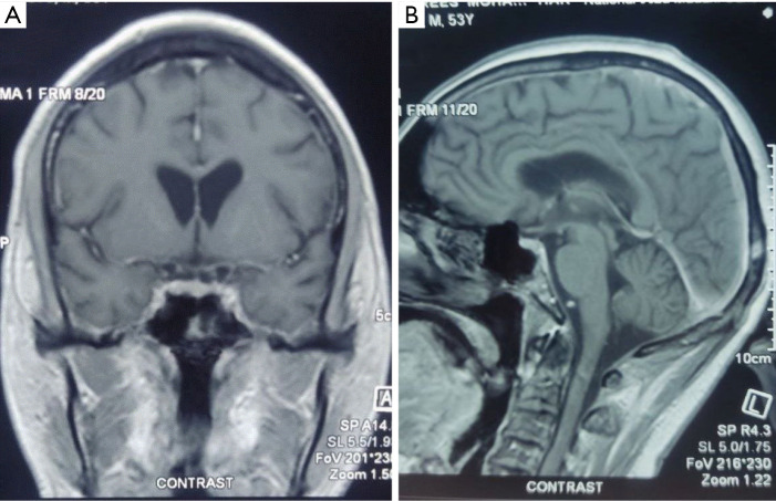
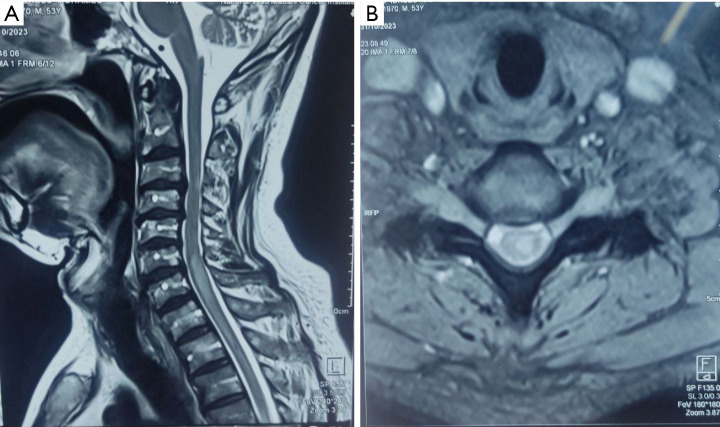
Case description: A 53-year-old hypertensive male, presented with subacute lower limbs weakness, sensory level below his nipples, and urine retention. Erythrocyte sedimentation rate (ESR) was above 112 mm/hour. Magnetic resonance imaging (MRI) spine showed a long segment of hyperintense signal seen on the T2-weighted image (T2WI) images in the spinal cord extending from C6 to D3 vertebral segments, with heterogeneous post-contrast enhancement. Cerebrospinal fluid (CSF) analysis showed lymphocytic pleocytosis with high protein and low glucose, and polymerase chain reaction (PCR) for Mycobacterium tuberculosis (MBTB) was positive. The patient received intravenous methylprednisolone daily for 5 days and standard anti-TB medications [rifampicin, isoniazid (INH), pyrazinamide, and ethambutol] for 12 months. However, repeated CSF analysis 3 months after starting anti-TB medications showed a negative PCR for MBTB, normal cell count, and glucose with slightly elevated protein. Still, the patient did not show any clinical improvement.
Conclusions: Long-segment tuberculous myelitis (LSTM) is a rare form of central nervous system TB that can be accompanied by tuberculous meningitis. It must be considered a differential diagnosis of neuromyelitis optica spectrum disorder (NMOSD), especially in endemic areas of TB, as the management approach is completely different.

 求助内容:
求助内容: 应助结果提醒方式:


