{"title":"A dual self-attentive transformer U-Net model for precise pancreatic segmentation and fat fraction estimation.","authors":"Ashok Shanmugam, Prianka Ramachandran Radhabai, Kavitha Kvn, Agbotiname Lucky Imoize","doi":"10.1186/s12880-025-01852-5","DOIUrl":null,"url":null,"abstract":"<p><p>Accurately segmenting the pancreas from abdominal computed tomography (CT) images is crucial for detecting and managing pancreatic diseases, such as diabetes and tumors. Type 2 diabetes and metabolic syndrome are associated with pancreatic fat accumulation. Calculating the fat fraction aids in the investigation of β-cell malfunction and insulin resistance. The most widely used pancreas segmentation technique is a U-shaped network based on deep convolutional neural networks (DCNNs). They struggle to capture long-range biases in an image because they rely on local receptive fields. This research proposes a novel dual Self-attentive Transformer Unet (DSTUnet) model for accurate pancreatic segmentation, addressing this problem. This model incorporates dual self-attention Swin transformers on both the encoder and decoder sides to facilitate global context extraction and refine candidate regions. After segmenting the pancreas using a DSTUnet, a histogram analysis is used to estimate the fat fraction. The suggested method demonstrated excellent performance on the standard dataset, achieving a DSC of 93.7% and an HD of 2.7 mm. The average volume of the pancreas was 92.42, and its fat volume fraction (FVF) was 13.37%.</p>","PeriodicalId":9020,"journal":{"name":"BMC Medical Imaging","volume":"25 1","pages":"315"},"PeriodicalIF":3.2000,"publicationDate":"2025-08-04","publicationTypes":"Journal Article","fieldsOfStudy":null,"isOpenAccess":false,"openAccessPdf":"https://www.ncbi.nlm.nih.gov/pmc/articles/PMC12323108/pdf/","citationCount":"0","resultStr":null,"platform":"Semanticscholar","paperid":null,"PeriodicalName":"BMC Medical Imaging","FirstCategoryId":"3","ListUrlMain":"https://doi.org/10.1186/s12880-025-01852-5","RegionNum":3,"RegionCategory":"医学","ArticlePicture":[],"TitleCN":null,"AbstractTextCN":null,"PMCID":null,"EPubDate":"","PubModel":"","JCR":"Q2","JCRName":"RADIOLOGY, NUCLEAR MEDICINE & MEDICAL IMAGING","Score":null,"Total":0}
引用次数: 0
Abstract
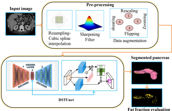
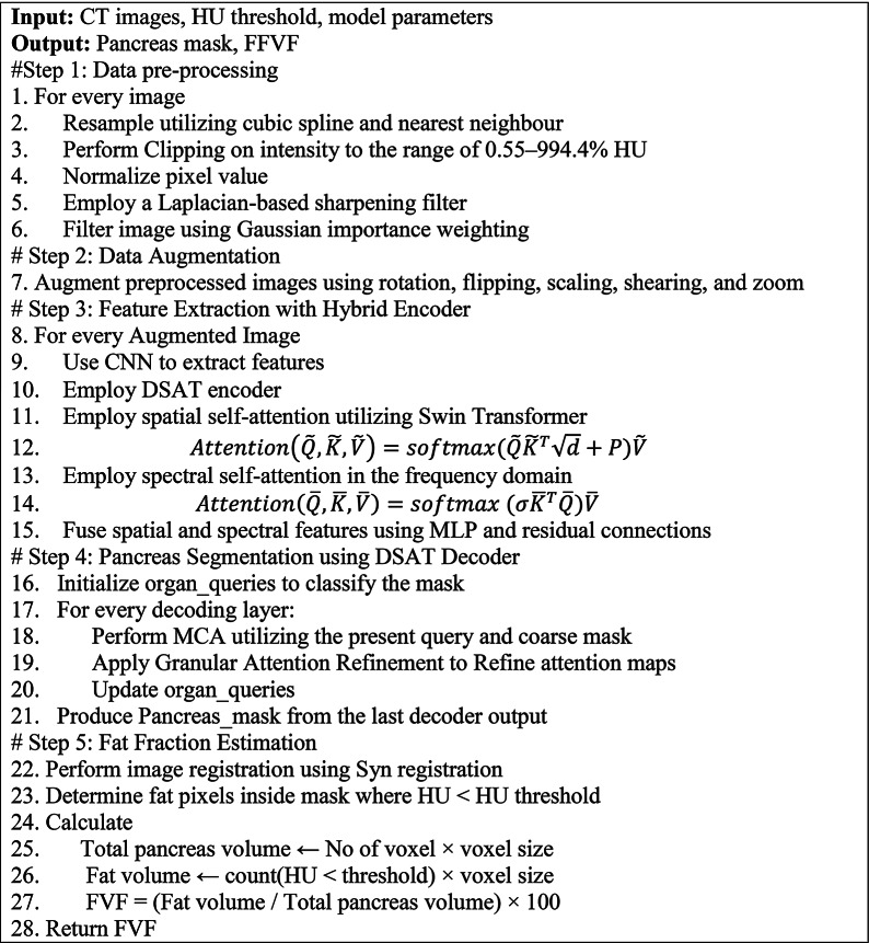
Accurately segmenting the pancreas from abdominal computed tomography (CT) images is crucial for detecting and managing pancreatic diseases, such as diabetes and tumors. Type 2 diabetes and metabolic syndrome are associated with pancreatic fat accumulation. Calculating the fat fraction aids in the investigation of β-cell malfunction and insulin resistance. The most widely used pancreas segmentation technique is a U-shaped network based on deep convolutional neural networks (DCNNs). They struggle to capture long-range biases in an image because they rely on local receptive fields. This research proposes a novel dual Self-attentive Transformer Unet (DSTUnet) model for accurate pancreatic segmentation, addressing this problem. This model incorporates dual self-attention Swin transformers on both the encoder and decoder sides to facilitate global context extraction and refine candidate regions. After segmenting the pancreas using a DSTUnet, a histogram analysis is used to estimate the fat fraction. The suggested method demonstrated excellent performance on the standard dataset, achieving a DSC of 93.7% and an HD of 2.7 mm. The average volume of the pancreas was 92.42, and its fat volume fraction (FVF) was 13.37%.
期刊介绍:
BMC Medical Imaging is an open access journal publishing original peer-reviewed research articles in the development, evaluation, and use of imaging techniques and image processing tools to diagnose and manage disease.

 求助内容:
求助内容: 应助结果提醒方式:


