Daniel Armbruster, Thomas Mueller, Wolfgang Driever
{"title":"Dopaminergic Neurons in the Zebrafish Subpallium Belong to the Extended Medial Amygdala","authors":"Daniel Armbruster, Thomas Mueller, Wolfgang Driever","doi":"10.1002/cne.70079","DOIUrl":null,"url":null,"abstract":"<p>The amygdala is a heterogeneous, multinuclear telencephalic structure critical for motivated and emotion-related behaviors in vertebrates. In ray-finned fish (Actinopterygii) like the teleost zebrafish, a telencephalic outward-growing process called eversion makes defining amygdaloid territories particularly challenging. Teleosts are also peculiar in that they develop numerous dopaminergic (DA) neurons in the subpallium, while in tetrapods, such populations are less prominent or appear only transiently. To shed light on the organization of the amygdala in teleosts, we pursued an evolutionary developmental approach focusing on the topological origin of subpallial DA neurons. Specifically, we analyzed the distribution of tyrosine hydroxylase (Th) in conjunction with expression patterns of <i>pax6a+b</i>, <i>isl1a</i>, <i>nkx2.1</i>, <i>lhx8a</i>, <i>otpa+b</i>, and <i>calb2a</i> as markers of different telencephalic subdivisions in brains of 5- and 30-day-old zebrafish (<i>Danio rerio</i>, Teleostei). Our data show that the previously identified dorsalmost division of the ventral telencephalon (Vdd) needs to be subdivided into an anteroventral <i>pax6a/b</i>-positive portion (Vdd1) and a posterodorsal <i>pax6a/b</i>-negative portion (Vdd2). This <i>pax6a</i>-negative Vdd2 portion develops into the extended medial amygdala (EMeA), including the DA population adjacent to the pallial–subpallial border. Our results also show that the EMeA DA neurons form a heterogeneous group of amygdaloid neurons because they differentially express <i>calb2a</i> and <i>sst7</i>. Our work sheds light on the early evolution and development of the amygdala and provides a foundation for functional analysis of the newly defined DA subtypes of the extended amygdala in zebrafish.</p>","PeriodicalId":15552,"journal":{"name":"Journal of Comparative Neurology","volume":"533 8","pages":""},"PeriodicalIF":2.1000,"publicationDate":"2025-08-05","publicationTypes":"Journal Article","fieldsOfStudy":null,"isOpenAccess":false,"openAccessPdf":"https://onlinelibrary.wiley.com/doi/epdf/10.1002/cne.70079","citationCount":"0","resultStr":null,"platform":"Semanticscholar","paperid":null,"PeriodicalName":"Journal of Comparative Neurology","FirstCategoryId":"3","ListUrlMain":"https://onlinelibrary.wiley.com/doi/10.1002/cne.70079","RegionNum":4,"RegionCategory":"医学","ArticlePicture":[],"TitleCN":null,"AbstractTextCN":null,"PMCID":null,"EPubDate":"","PubModel":"","JCR":"Q3","JCRName":"NEUROSCIENCES","Score":null,"Total":0}
引用次数: 0
Abstract
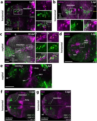
The amygdala is a heterogeneous, multinuclear telencephalic structure critical for motivated and emotion-related behaviors in vertebrates. In ray-finned fish (Actinopterygii) like the teleost zebrafish, a telencephalic outward-growing process called eversion makes defining amygdaloid territories particularly challenging. Teleosts are also peculiar in that they develop numerous dopaminergic (DA) neurons in the subpallium, while in tetrapods, such populations are less prominent or appear only transiently. To shed light on the organization of the amygdala in teleosts, we pursued an evolutionary developmental approach focusing on the topological origin of subpallial DA neurons. Specifically, we analyzed the distribution of tyrosine hydroxylase (Th) in conjunction with expression patterns of pax6a+b, isl1a, nkx2.1, lhx8a, otpa+b, and calb2a as markers of different telencephalic subdivisions in brains of 5- and 30-day-old zebrafish (Danio rerio, Teleostei). Our data show that the previously identified dorsalmost division of the ventral telencephalon (Vdd) needs to be subdivided into an anteroventral pax6a/b-positive portion (Vdd1) and a posterodorsal pax6a/b-negative portion (Vdd2). This pax6a-negative Vdd2 portion develops into the extended medial amygdala (EMeA), including the DA population adjacent to the pallial–subpallial border. Our results also show that the EMeA DA neurons form a heterogeneous group of amygdaloid neurons because they differentially express calb2a and sst7. Our work sheds light on the early evolution and development of the amygdala and provides a foundation for functional analysis of the newly defined DA subtypes of the extended amygdala in zebrafish.
期刊介绍:
Established in 1891, JCN is the oldest continually published basic neuroscience journal. Historically, as the name suggests, the journal focused on a comparison among species to uncover the intricacies of how the brain functions. In modern times, this research is called systems neuroscience where animal models are used to mimic core cognitive processes with the ultimate goal of understanding neural circuits and connections that give rise to behavioral patterns and different neural states.
Research published in JCN covers all species from invertebrates to humans, and the reports inform the readers about the function and organization of nervous systems in species with an emphasis on the way that species adaptations inform about the function or organization of the nervous systems, rather than on their evolution per se.
JCN publishes primary research articles and critical commentaries and review-type articles offering expert insight in to cutting edge research in the field of systems neuroscience; a complete list of contribution types is given in the Author Guidelines. For primary research contributions, only full-length investigative reports are desired; the journal does not accept short communications.

 求助内容:
求助内容: 应助结果提醒方式:


