Kaige Wang, Linhui Yang, Zhen Kang, Zhuang Luo, Dan Liu, Fen Tan, Weimin Li
{"title":"Clinical characteristics of pulmonary sarcoidosis in China: a retrospective, multicenter study.","authors":"Kaige Wang, Linhui Yang, Zhen Kang, Zhuang Luo, Dan Liu, Fen Tan, Weimin Li","doi":"10.1080/07853890.2025.2540017","DOIUrl":null,"url":null,"abstract":"<p><strong>Background: </strong>Patients with pulmonary sarcoidosis or intrathoracic lymph node tuberculosis (TB) may present with comparable clinical manifestations that pose challenges in differentiation. This study aims to improve the diagnostic accuracy of pulmonary sarcoidosis.</p><p><strong>Methods: </strong>A retrospective analysis of patients diagnosed with pulmonary sarcoidosis or intrathoracic lymph node TB within the past decade at four tertiary hospitals in China was conducted. According to the inclusion and exclusion criteria, a total of 968 patients were ultimately enrolled in the study, comprising 477 individuals diagnosed with pulmonary sarcoidosis and 491 individuals diagnosed with intrathoracic lymph node TB. The analysis focused on general information, clinical manifestations, and auxiliary examination results, with a comparative analysis between the two groups.</p><p><strong>Results: </strong>The median age of onset for pulmonary sarcoidosis was 50 years, with females accounting for 68.94% of the patients. Common symptoms of pulmonary sarcoidosis included cough, sputum production, dyspnea, and chest pain, while approximately 34.12% of patients were asymptomatic. Fever, fatigue, and night sweats occurred less frequently in pulmonary sarcoidosis patients than in those with intrathoracic lymph node TB. Uveitis and myocardial sarcoidosis were observed exclusively in pulmonary sarcoidosis patients. The median time from symptom onset to the diagnosis of pulmonary sarcoidosis was up to three months. Approximately 47.29% of pulmonary sarcoidosis patients had reduced peripheral blood lymphocyte counts, and 94.12% exhibited symmetric enlargement of hilar lymph nodes on chest CT. Both pulmonary sarcoidosis and intrathoracic lymph node TB showed granulomatous inflammation, with 64.36% of intrathoracic lymph node TB cases presenting necrotic foci. Bronchoscopy was the primary method for biopsy, and only 11.06% of pulmonary sarcoidosis patients had multiple nodules in the tracheal or bronchial mucosa, with a low positivity rate for pathogen tests.</p><p><strong>Conclusion: </strong>Pulmonary sarcoidosis predominantly affects middle-aged and young women and can be differentiated from intrathoracic lymph node TB by the presence of uveitis and myocardial sarcoidosis, although these manifestations are rare. A significant proportion of pulmonary sarcoidosis patients experience a reduction in their peripheral blood lymphocyte count. Chest CT scans often reveal symmetric bilateral enlargement of hilar lymph nodes, and in some cases, multiple nodules in the tracheal or bronchial mucosa. Both pulmonary sarcoidosis and intrathoracic lymph node TB show granulomatous inflammation, but tuberculosis lesions are more likely to necrose.</p>","PeriodicalId":93874,"journal":{"name":"Annals of medicine","volume":"57 1","pages":"2540017"},"PeriodicalIF":4.3000,"publicationDate":"2025-12-01","publicationTypes":"Journal Article","fieldsOfStudy":null,"isOpenAccess":false,"openAccessPdf":"https://www.ncbi.nlm.nih.gov/pmc/articles/PMC12322989/pdf/","citationCount":"0","resultStr":null,"platform":"Semanticscholar","paperid":null,"PeriodicalName":"Annals of medicine","FirstCategoryId":"1085","ListUrlMain":"https://doi.org/10.1080/07853890.2025.2540017","RegionNum":0,"RegionCategory":null,"ArticlePicture":[],"TitleCN":null,"AbstractTextCN":null,"PMCID":null,"EPubDate":"2025/8/3 0:00:00","PubModel":"Epub","JCR":"","JCRName":"","Score":null,"Total":0}
引用次数: 0
Abstract
Background: Patients with pulmonary sarcoidosis or intrathoracic lymph node tuberculosis (TB) may present with comparable clinical manifestations that pose challenges in differentiation. This study aims to improve the diagnostic accuracy of pulmonary sarcoidosis.
Methods: A retrospective analysis of patients diagnosed with pulmonary sarcoidosis or intrathoracic lymph node TB within the past decade at four tertiary hospitals in China was conducted. According to the inclusion and exclusion criteria, a total of 968 patients were ultimately enrolled in the study, comprising 477 individuals diagnosed with pulmonary sarcoidosis and 491 individuals diagnosed with intrathoracic lymph node TB. The analysis focused on general information, clinical manifestations, and auxiliary examination results, with a comparative analysis between the two groups.
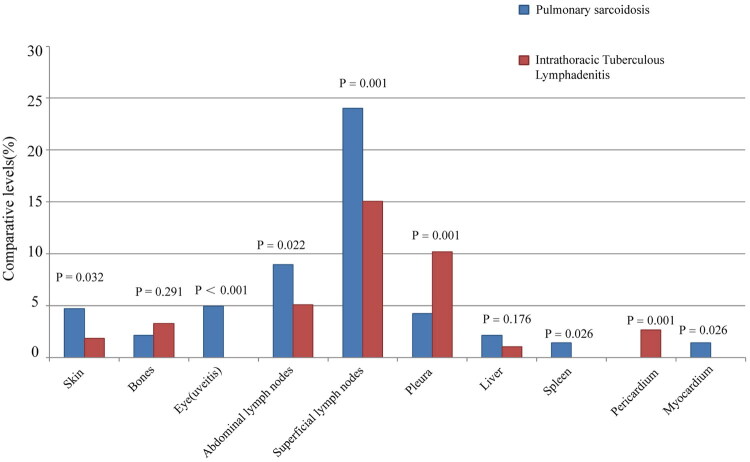
Results: The median age of onset for pulmonary sarcoidosis was 50 years, with females accounting for 68.94% of the patients. Common symptoms of pulmonary sarcoidosis included cough, sputum production, dyspnea, and chest pain, while approximately 34.12% of patients were asymptomatic. Fever, fatigue, and night sweats occurred less frequently in pulmonary sarcoidosis patients than in those with intrathoracic lymph node TB. Uveitis and myocardial sarcoidosis were observed exclusively in pulmonary sarcoidosis patients. The median time from symptom onset to the diagnosis of pulmonary sarcoidosis was up to three months. Approximately 47.29% of pulmonary sarcoidosis patients had reduced peripheral blood lymphocyte counts, and 94.12% exhibited symmetric enlargement of hilar lymph nodes on chest CT. Both pulmonary sarcoidosis and intrathoracic lymph node TB showed granulomatous inflammation, with 64.36% of intrathoracic lymph node TB cases presenting necrotic foci. Bronchoscopy was the primary method for biopsy, and only 11.06% of pulmonary sarcoidosis patients had multiple nodules in the tracheal or bronchial mucosa, with a low positivity rate for pathogen tests.
Conclusion: Pulmonary sarcoidosis predominantly affects middle-aged and young women and can be differentiated from intrathoracic lymph node TB by the presence of uveitis and myocardial sarcoidosis, although these manifestations are rare. A significant proportion of pulmonary sarcoidosis patients experience a reduction in their peripheral blood lymphocyte count. Chest CT scans often reveal symmetric bilateral enlargement of hilar lymph nodes, and in some cases, multiple nodules in the tracheal or bronchial mucosa. Both pulmonary sarcoidosis and intrathoracic lymph node TB show granulomatous inflammation, but tuberculosis lesions are more likely to necrose.

 求助内容:
求助内容: 应助结果提醒方式:


