Tianjie Zhang, Yan Liu, Mengmei Wang, Miao He, Min Yu, Yi Li, Ye Song
{"title":"Diaphragm Assessment by Multimodal Ultrasound Imaging in Patients with Chronic Obstructive Pulmonary Disease: A Prospective Observational Study.","authors":"Tianjie Zhang, Yan Liu, Mengmei Wang, Miao He, Min Yu, Yi Li, Ye Song","doi":"10.2147/COPD.S527119","DOIUrl":null,"url":null,"abstract":"<p><strong>Background: </strong>Chronic obstructive pulmonary disease (COPD) patients often exhibit diaphragmatic dysfunction which can be effectively assessed using ultrasonography. This study aims to evaluate diaphragmatic function in COPD patients through multimodal ultrasound imaging and to identify key parameters.</p><p><strong>Methods: </strong>This study consecutively enrolled 75 COPD patients and 75 healthy subjects. Measurements of diaphragm contraction, motion-related parameters and tissue Doppler imaging (TDI) parameters were conducted and recorded. Clinically relevant data were collected. Baseline demographics, spirometry results, and ultrasound data were compared between COPD patients and healthy subjects. Receiver Operating Characteristic (ROC) curve was constructed to evaluate the diagnostic value of diaphragmatic ultrasound parameters for COPD patients.</p><p><strong>Results: </strong>Diaphragm at the end of tidal inspiration (DT_insp), diaphragm thickening fraction (DTF), diaphragmatic excursion during deep breathing (DE_DB) were significantly lower in COPD patients than in healthy subjects, conversely diaphragmatic excursion during quiet breathing (DE_QB), diaphragmatic contraction velocity during quiet breathing (DCV_QB), peak contraction velocity(PCV), peak relaxation velocity (PRV), velocity-time integral (VTI) were higher in COPD patients than in healthy subjects. DT_insp, DTF, DE_DB values decreased as COPD severity increased, conversely, DE_QB, DCV_QB, PCV, PRV and VTI exhibited an upward trend with COPD severity. DTF was positively correlated with FEV1 predicted (r=0.713, P=0.000), DE_QB (r=-0.740 and -0.889), PCV (r=-0.609 and -0.778), PRV (r=-0.686 and -0.857) were negatively correlated with FEV1/FVC and FEV1 predicted (P=0.000). Meanwhile, DE_QB, DCV_QB, PCV and PRV exhibited superior performance in predicting COPD, with AUC values of 0.906, 0.833, 0.859 and 0.833, respectively. DE_QB exhibited 81.33% sensitivity, while DTF, DE_QB, DE_DB, PCV and PRV showed high specificity (98.67%, 90.67%, 96.00%, 97.33% and 100%, respectively).</p><p><strong>Conclusion: </strong>Multimodal ultrasound imaging offers a sensitive approach for detecting diaphragmatic dysfunction in COPD patients. Diaphragm ultrasound parameters correlate with pulmonary function and COPD severity, indicating that these parameters can provide valuable insights into disease progression and management.</p>","PeriodicalId":48818,"journal":{"name":"International Journal of Chronic Obstructive Pulmonary Disease","volume":"20 ","pages":"2629-2638"},"PeriodicalIF":3.1000,"publicationDate":"2025-07-29","publicationTypes":"Journal Article","fieldsOfStudy":null,"isOpenAccess":false,"openAccessPdf":"https://www.ncbi.nlm.nih.gov/pmc/articles/PMC12317707/pdf/","citationCount":"0","resultStr":null,"platform":"Semanticscholar","paperid":null,"PeriodicalName":"International Journal of Chronic Obstructive Pulmonary Disease","FirstCategoryId":"3","ListUrlMain":"https://doi.org/10.2147/COPD.S527119","RegionNum":3,"RegionCategory":"医学","ArticlePicture":[],"TitleCN":null,"AbstractTextCN":null,"PMCID":null,"EPubDate":"2025/1/1 0:00:00","PubModel":"eCollection","JCR":"Q2","JCRName":"RESPIRATORY SYSTEM","Score":null,"Total":0}
引用次数: 0
Abstract
Background: Chronic obstructive pulmonary disease (COPD) patients often exhibit diaphragmatic dysfunction which can be effectively assessed using ultrasonography. This study aims to evaluate diaphragmatic function in COPD patients through multimodal ultrasound imaging and to identify key parameters.
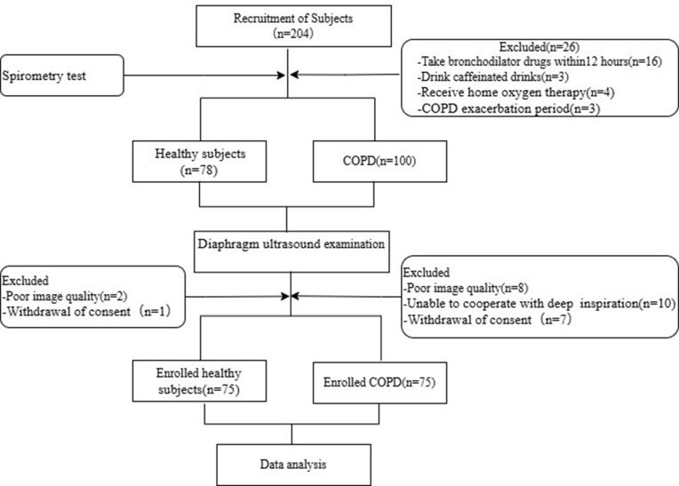
Methods: This study consecutively enrolled 75 COPD patients and 75 healthy subjects. Measurements of diaphragm contraction, motion-related parameters and tissue Doppler imaging (TDI) parameters were conducted and recorded. Clinically relevant data were collected. Baseline demographics, spirometry results, and ultrasound data were compared between COPD patients and healthy subjects. Receiver Operating Characteristic (ROC) curve was constructed to evaluate the diagnostic value of diaphragmatic ultrasound parameters for COPD patients.
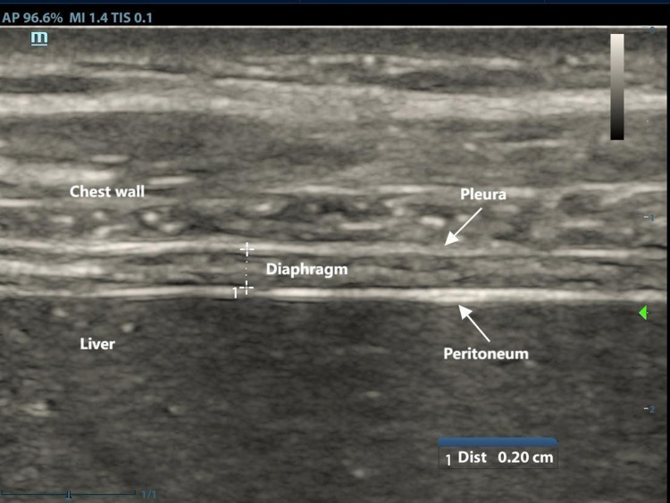
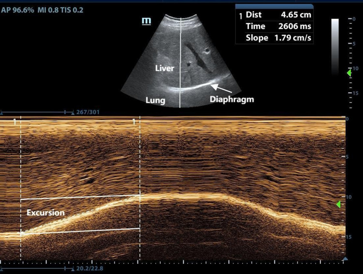
Results: Diaphragm at the end of tidal inspiration (DT_insp), diaphragm thickening fraction (DTF), diaphragmatic excursion during deep breathing (DE_DB) were significantly lower in COPD patients than in healthy subjects, conversely diaphragmatic excursion during quiet breathing (DE_QB), diaphragmatic contraction velocity during quiet breathing (DCV_QB), peak contraction velocity(PCV), peak relaxation velocity (PRV), velocity-time integral (VTI) were higher in COPD patients than in healthy subjects. DT_insp, DTF, DE_DB values decreased as COPD severity increased, conversely, DE_QB, DCV_QB, PCV, PRV and VTI exhibited an upward trend with COPD severity. DTF was positively correlated with FEV1 predicted (r=0.713, P=0.000), DE_QB (r=-0.740 and -0.889), PCV (r=-0.609 and -0.778), PRV (r=-0.686 and -0.857) were negatively correlated with FEV1/FVC and FEV1 predicted (P=0.000). Meanwhile, DE_QB, DCV_QB, PCV and PRV exhibited superior performance in predicting COPD, with AUC values of 0.906, 0.833, 0.859 and 0.833, respectively. DE_QB exhibited 81.33% sensitivity, while DTF, DE_QB, DE_DB, PCV and PRV showed high specificity (98.67%, 90.67%, 96.00%, 97.33% and 100%, respectively).
Conclusion: Multimodal ultrasound imaging offers a sensitive approach for detecting diaphragmatic dysfunction in COPD patients. Diaphragm ultrasound parameters correlate with pulmonary function and COPD severity, indicating that these parameters can provide valuable insights into disease progression and management.
期刊介绍:
An international, peer-reviewed journal of therapeutics and pharmacology focusing on concise rapid reporting of clinical studies and reviews in COPD. Special focus will be given to the pathophysiological processes underlying the disease, intervention programs, patient focused education, and self management protocols. This journal is directed at specialists and healthcare professionals
 求助内容:
求助内容: 应助结果提醒方式:


