Sayed Shojaedin Shayegh, Zahra Mohammadi, Mohammad Amin Bafandeh, Mohammad Reza Nazemalroaya
{"title":"The Use of an Intraoral Scanner for the Fabrication of Maxillary Obturator Prosthesis in a Young Adult With Oronasal Fistula: A Case Report.","authors":"Sayed Shojaedin Shayegh, Zahra Mohammadi, Mohammad Amin Bafandeh, Mohammad Reza Nazemalroaya","doi":"10.1155/crid/1167521","DOIUrl":null,"url":null,"abstract":"<p><p><b>Background:</b> Cleft lip and palate require complex treatment, often involving early surgery. Postoperative complications, such as palatal fistulas, can impair speech and feeding. While surgical correction is standard, large fistulas may pose challenges due to age, cost, and recurrence risks. Obturator prostheses provide a nonsurgical alternative, but digital impression techniques for their fabrication are underutilized. This case report explores intraoral digital impressions for creating obturator/speech aid appliances in a patient with cleft palate deformities. <b>Methods:</b> A 17-year-old female with Class III malocclusion on a Class III skeletal base and increased facial proportion and IOFTN score of 5, with cleft palate-related eating difficulties, missing anterior teeth, and worn dentition, underwent intraoral scanning. Digital files were used to fabricate a premolar-to-premolar obturator, with relief areas and teeth arranged on printed casts. <b>Results:</b> The appliance effectively addressed functional and aesthetic concerns. <b>Conclusion:</b> Digital impressions offer precise, efficient, and comfortable fabrication of obturator prostheses compared to conventional methods. Despite initial costs, they reduce chair time, enhance accuracy for dental hard tissues, and improve patient experience, particularly for young patients with cleft lip and palate.</p>","PeriodicalId":46841,"journal":{"name":"Case Reports in Dentistry","volume":"2025 ","pages":"1167521"},"PeriodicalIF":0.9000,"publicationDate":"2025-07-26","publicationTypes":"Journal Article","fieldsOfStudy":null,"isOpenAccess":false,"openAccessPdf":"https://www.ncbi.nlm.nih.gov/pmc/articles/PMC12317820/pdf/","citationCount":"0","resultStr":null,"platform":"Semanticscholar","paperid":null,"PeriodicalName":"Case Reports in Dentistry","FirstCategoryId":"1085","ListUrlMain":"https://doi.org/10.1155/crid/1167521","RegionNum":0,"RegionCategory":null,"ArticlePicture":[],"TitleCN":null,"AbstractTextCN":null,"PMCID":null,"EPubDate":"2025/1/1 0:00:00","PubModel":"eCollection","JCR":"Q4","JCRName":"DENTISTRY, ORAL SURGERY & MEDICINE","Score":null,"Total":0}
引用次数: 0
Abstract
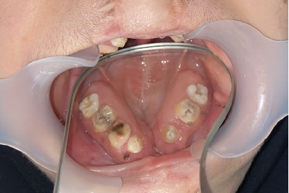
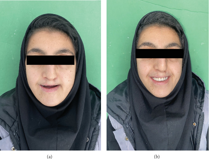
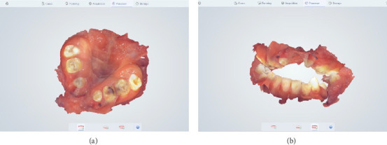
Background: Cleft lip and palate require complex treatment, often involving early surgery. Postoperative complications, such as palatal fistulas, can impair speech and feeding. While surgical correction is standard, large fistulas may pose challenges due to age, cost, and recurrence risks. Obturator prostheses provide a nonsurgical alternative, but digital impression techniques for their fabrication are underutilized. This case report explores intraoral digital impressions for creating obturator/speech aid appliances in a patient with cleft palate deformities. Methods: A 17-year-old female with Class III malocclusion on a Class III skeletal base and increased facial proportion and IOFTN score of 5, with cleft palate-related eating difficulties, missing anterior teeth, and worn dentition, underwent intraoral scanning. Digital files were used to fabricate a premolar-to-premolar obturator, with relief areas and teeth arranged on printed casts. Results: The appliance effectively addressed functional and aesthetic concerns. Conclusion: Digital impressions offer precise, efficient, and comfortable fabrication of obturator prostheses compared to conventional methods. Despite initial costs, they reduce chair time, enhance accuracy for dental hard tissues, and improve patient experience, particularly for young patients with cleft lip and palate.
期刊介绍:
Case Reports in Dentistry is a peer-reviewed, Open Access journal that publishes case reports and case series in all areas of dentistry, including periodontal diseases, dental implants, oral pathology, as well as oral and maxillofacial surgery.

 求助内容:
求助内容: 应助结果提醒方式:


