{"title":"Guided Endodontic Technique in Mandibular Incisors with Pulp Canal Obliteration: A Case Report.","authors":"Saide Nabavi, Erfan Latifian","doi":"10.22037/iej.v20i1.47737","DOIUrl":null,"url":null,"abstract":"<p><p>The American Association of Endodontics classifies the management of pulp canal obliteration (PCO) cases as a high-risk difficulty. This classification indicates an increased likelihood of procedural errors. However, advancements in dental technology have simplified the endodontic treatment of calcified teeth. Guided endodontics uses cone-beam computed tomography (CBCT) and a three-dimensional (3D) printer to create a patient-specific guide, enhancing accuracy and predictability in complex cases. A healthy 60-year-old male was referred by a prosthodontist for root canal treatment on teeth #23, #24, and #25. Periapical radiographic examination revealed partial PCO in all teeth. After choosing guided endodontics treatment, CBCT imaging and intraoral 3D scanning were utilized to design a patient-specific endodontic guide. After verifying the stability of the endo-guide, access cavities were prepared using a Munce Discovery bur. Following the negotiation of the canals, the working length was determined. Root canal treatment was performed using rotary files up to size 25/0.04% and sodium hypochlorite irrigation. The obturation was completed using the warm condensation technique. Although PCO was present, no complications occurred during treatment. The teeth remained completely asymptomatic and functional, demonstrating the success of the treatment. Guided endodontics can be a practical and predictable approach for managing PCO in mandibular incisors. This technique provides accurate canal location, reduces procedural errors, and preserves tooth structure. Despite concerns about cost, radiation exposure, and challenges with anatomical variations, it represents a promising advancement in endodontic treatment.</p>","PeriodicalId":14534,"journal":{"name":"Iranian Endodontic Journal","volume":"20 1","pages":"e21"},"PeriodicalIF":0.0000,"publicationDate":"2025-01-01","publicationTypes":"Journal Article","fieldsOfStudy":null,"isOpenAccess":false,"openAccessPdf":"https://www.ncbi.nlm.nih.gov/pmc/articles/PMC12318319/pdf/","citationCount":"0","resultStr":null,"platform":"Semanticscholar","paperid":null,"PeriodicalName":"Iranian Endodontic Journal","FirstCategoryId":"1085","ListUrlMain":"https://doi.org/10.22037/iej.v20i1.47737","RegionNum":0,"RegionCategory":null,"ArticlePicture":[],"TitleCN":null,"AbstractTextCN":null,"PMCID":null,"EPubDate":"2025/5/11 0:00:00","PubModel":"Epub","JCR":"Q3","JCRName":"Dentistry","Score":null,"Total":0}
引用次数: 0
Abstract
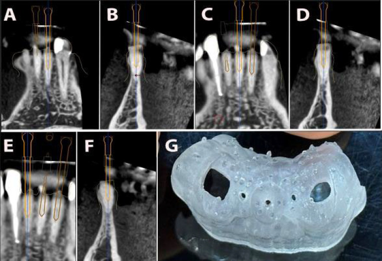
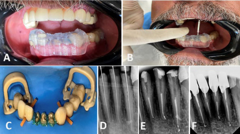
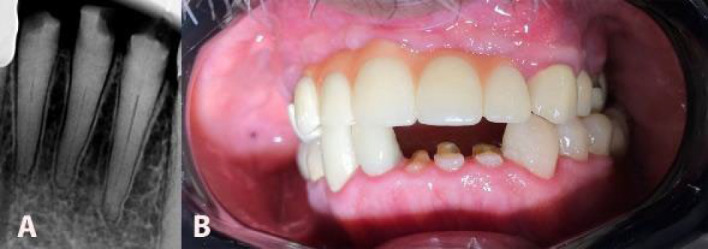
The American Association of Endodontics classifies the management of pulp canal obliteration (PCO) cases as a high-risk difficulty. This classification indicates an increased likelihood of procedural errors. However, advancements in dental technology have simplified the endodontic treatment of calcified teeth. Guided endodontics uses cone-beam computed tomography (CBCT) and a three-dimensional (3D) printer to create a patient-specific guide, enhancing accuracy and predictability in complex cases. A healthy 60-year-old male was referred by a prosthodontist for root canal treatment on teeth #23, #24, and #25. Periapical radiographic examination revealed partial PCO in all teeth. After choosing guided endodontics treatment, CBCT imaging and intraoral 3D scanning were utilized to design a patient-specific endodontic guide. After verifying the stability of the endo-guide, access cavities were prepared using a Munce Discovery bur. Following the negotiation of the canals, the working length was determined. Root canal treatment was performed using rotary files up to size 25/0.04% and sodium hypochlorite irrigation. The obturation was completed using the warm condensation technique. Although PCO was present, no complications occurred during treatment. The teeth remained completely asymptomatic and functional, demonstrating the success of the treatment. Guided endodontics can be a practical and predictable approach for managing PCO in mandibular incisors. This technique provides accurate canal location, reduces procedural errors, and preserves tooth structure. Despite concerns about cost, radiation exposure, and challenges with anatomical variations, it represents a promising advancement in endodontic treatment.
期刊介绍:
The Iranian Endodontic Journal (IEJ) is an international peer-reviewed biomedical publication, the aim of which is to provide a scientific medium of communication for researchers throughout the globe. IEJ aims to publish the highest quality articles, both clinical and scientific, on all aspects of Endodontics. The journal is an official Journal of the Iranian Center for Endodontic Research (ICER) and the Iranian Association of Endodontists (IAE). The Journal welcomes articles related to the scientific or applied aspects of endodontics e.g. original researches, systematic reviews, meta-analyses, review articles, clinical trials, case series/reports, hypotheses, letters to the editor, etc. From the beginning (i.e. since 2006), the IEJ was the first open access endodontic journal in the world, which gave readers free and instant access to published articles and enabling them faster discovery of the latest endodontic research.

 求助内容:
求助内容: 应助结果提醒方式:


