Zixuan Meng, Yuehua Han, Linda Ruan, Chenming Xu, Mengxiao Zhang, Hao Liu
{"title":"Establishment and Evaluation of HepG2 Cell Insulin Resistance Model.","authors":"Zixuan Meng, Yuehua Han, Linda Ruan, Chenming Xu, Mengxiao Zhang, Hao Liu","doi":"10.2147/DMSO.S523821","DOIUrl":null,"url":null,"abstract":"<p><strong>Objective: </strong>Establishing HepG2 cell insulin resistance models using glucosamine, high glucose with high insulin and palmitic acid and briefly evaluating them to provide reliable models for insulin resistance research.</p><p><strong>Methods: </strong>Three methods were used to induce insulin resistance in HepG2 cells, and glucose uptake and consumption, glucose metabolism-related mRNA and p-AKT/AKT protein levels and RNA-seq were detected to compare the three induction methods.</p><p><strong>Results: </strong>Glucose consumption capacity was reduced after glucosamine and palmitic acid induction and did not change significantly after high glucose with high insulin induction. Glucose uptake capacity was not significantly changed after glucosamine and high glucose with high insulin induction and was reduced after palmitic acid induction. After high insulin stimulation, p-AKT/AKT levels were elevated in glucosamine and high glucose with high insulin induction and did not change significantly in palmitic acid induction. <i>G6pase</i>, <i>PC</i>, and <i>PCK1</i> were elevated after glucosamine and palmitic acid stimulation, and only PCK1 was elevated after high glucose with high insulin stimulation. The transcriptomes of cells induced by the three methods differed widely.</p><p><strong>Conclusion: </strong>Treatment with 0.2 mM palmitic acid for 24 h is a simple and stable method to induce insulin resistance in HepG2 cells.</p>","PeriodicalId":11116,"journal":{"name":"Diabetes, Metabolic Syndrome and Obesity: Targets and Therapy","volume":"18 ","pages":"2573-2584"},"PeriodicalIF":3.0000,"publicationDate":"2025-07-29","publicationTypes":"Journal Article","fieldsOfStudy":null,"isOpenAccess":false,"openAccessPdf":"https://www.ncbi.nlm.nih.gov/pmc/articles/PMC12317709/pdf/","citationCount":"0","resultStr":null,"platform":"Semanticscholar","paperid":null,"PeriodicalName":"Diabetes, Metabolic Syndrome and Obesity: Targets and Therapy","FirstCategoryId":"3","ListUrlMain":"https://doi.org/10.2147/DMSO.S523821","RegionNum":3,"RegionCategory":"医学","ArticlePicture":[],"TitleCN":null,"AbstractTextCN":null,"PMCID":null,"EPubDate":"2025/1/1 0:00:00","PubModel":"eCollection","JCR":"Q3","JCRName":"ENDOCRINOLOGY & METABOLISM","Score":null,"Total":0}
引用次数: 0
Abstract
Objective: Establishing HepG2 cell insulin resistance models using glucosamine, high glucose with high insulin and palmitic acid and briefly evaluating them to provide reliable models for insulin resistance research.
Methods: Three methods were used to induce insulin resistance in HepG2 cells, and glucose uptake and consumption, glucose metabolism-related mRNA and p-AKT/AKT protein levels and RNA-seq were detected to compare the three induction methods.
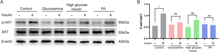
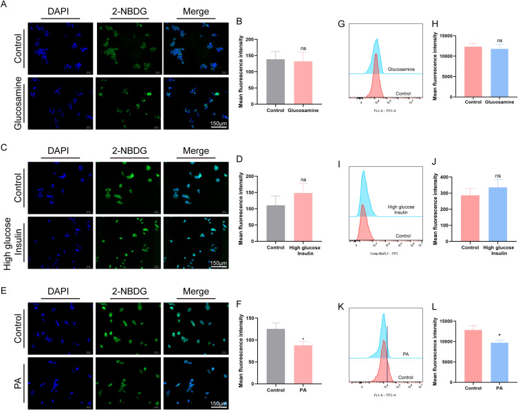
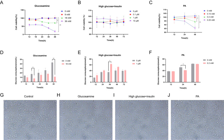
Results: Glucose consumption capacity was reduced after glucosamine and palmitic acid induction and did not change significantly after high glucose with high insulin induction. Glucose uptake capacity was not significantly changed after glucosamine and high glucose with high insulin induction and was reduced after palmitic acid induction. After high insulin stimulation, p-AKT/AKT levels were elevated in glucosamine and high glucose with high insulin induction and did not change significantly in palmitic acid induction. G6pase, PC, and PCK1 were elevated after glucosamine and palmitic acid stimulation, and only PCK1 was elevated after high glucose with high insulin stimulation. The transcriptomes of cells induced by the three methods differed widely.
Conclusion: Treatment with 0.2 mM palmitic acid for 24 h is a simple and stable method to induce insulin resistance in HepG2 cells.
期刊介绍:
An international, peer-reviewed, open access, online journal. The journal is committed to the rapid publication of the latest laboratory and clinical findings in the fields of diabetes, metabolic syndrome and obesity research. Original research, review, case reports, hypothesis formation, expert opinion and commentaries are all considered for publication.
 求助内容:
求助内容: 应助结果提醒方式:


