Sanjeeta Sitaula, Chiranjiwi Shah, Ganga Sagar Shah, Rajeev Ojha
{"title":"Neuro-Ophthalmic Manifestation Associated With COVID-19 in a Tertiary Eye Center in Nepal.","authors":"Sanjeeta Sitaula, Chiranjiwi Shah, Ganga Sagar Shah, Rajeev Ojha","doi":"10.1155/crop/6694537","DOIUrl":null,"url":null,"abstract":"<p><p>The coronavirus disease 2019 (COVID-19) primarily involves the respiratory system, but can manifest with a variety of neuro-ophthalmic symptoms. Here, we describe three cases presenting with neuro-ophthalmic manifestations secondary to COVID-19 at a tertiary center in Nepal. The first case was a 42-year-old male with sudden onset painless loss of vision noticed in the right eye (RE) after COVID-19 infection. Examination findings in the RE showed best corrected visual acuity (BCVA) of 6/18 with relative afferent pupillary defect positive and superior sectoral disk edema in the same eye. The case was diagnosed as RE nonarteritic ischemic optic neuropathy associated with COVID-19 infection. Our second case was a 41-year-old female who developed bilateral sudden diminution of vision associated with headache and vomiting on the third day of testing positive for COVID-19 infection. She had bilateral BCVA of 6/12 and sluggishly reacting pupils in both eyes. Dilated fundus examination showed established disk edema. Imaging of the brain showed dural venous sinus (transverse and sagittal) thrombosis. So, the diagnosis of papilledema secondary to COVID-19 associated cerebral venous sinus thrombosis (CVST) was established. The third case was a 40-year-old male with right sixth cranial nerve palsy, ischemic stroke involving the right occipital lobe and posterior limb of the right internal capsule along with thrombosis of the left common iliac artery in the absence of any other preexisting vascular risk factors. Severe inflammatory reaction to COVID-19 causing a hypercoagulable state may be the causal factor in neuro-ophthalmic findings in our case series.</p>","PeriodicalId":9603,"journal":{"name":"Case Reports in Ophthalmological Medicine","volume":"2025 ","pages":"6694537"},"PeriodicalIF":0.4000,"publicationDate":"2025-07-26","publicationTypes":"Journal Article","fieldsOfStudy":null,"isOpenAccess":false,"openAccessPdf":"https://www.ncbi.nlm.nih.gov/pmc/articles/PMC12317809/pdf/","citationCount":"0","resultStr":null,"platform":"Semanticscholar","paperid":null,"PeriodicalName":"Case Reports in Ophthalmological Medicine","FirstCategoryId":"1085","ListUrlMain":"https://doi.org/10.1155/crop/6694537","RegionNum":0,"RegionCategory":null,"ArticlePicture":[],"TitleCN":null,"AbstractTextCN":null,"PMCID":null,"EPubDate":"2025/1/1 0:00:00","PubModel":"eCollection","JCR":"Q4","JCRName":"OPHTHALMOLOGY","Score":null,"Total":0}
引用次数: 0
Abstract
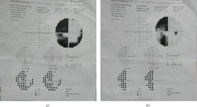
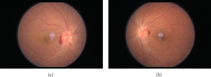
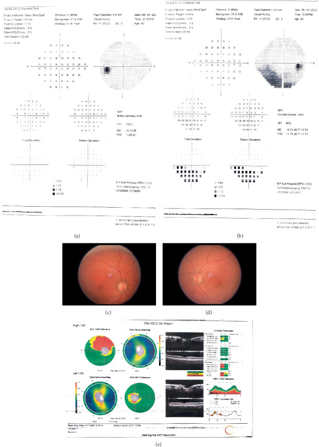
The coronavirus disease 2019 (COVID-19) primarily involves the respiratory system, but can manifest with a variety of neuro-ophthalmic symptoms. Here, we describe three cases presenting with neuro-ophthalmic manifestations secondary to COVID-19 at a tertiary center in Nepal. The first case was a 42-year-old male with sudden onset painless loss of vision noticed in the right eye (RE) after COVID-19 infection. Examination findings in the RE showed best corrected visual acuity (BCVA) of 6/18 with relative afferent pupillary defect positive and superior sectoral disk edema in the same eye. The case was diagnosed as RE nonarteritic ischemic optic neuropathy associated with COVID-19 infection. Our second case was a 41-year-old female who developed bilateral sudden diminution of vision associated with headache and vomiting on the third day of testing positive for COVID-19 infection. She had bilateral BCVA of 6/12 and sluggishly reacting pupils in both eyes. Dilated fundus examination showed established disk edema. Imaging of the brain showed dural venous sinus (transverse and sagittal) thrombosis. So, the diagnosis of papilledema secondary to COVID-19 associated cerebral venous sinus thrombosis (CVST) was established. The third case was a 40-year-old male with right sixth cranial nerve palsy, ischemic stroke involving the right occipital lobe and posterior limb of the right internal capsule along with thrombosis of the left common iliac artery in the absence of any other preexisting vascular risk factors. Severe inflammatory reaction to COVID-19 causing a hypercoagulable state may be the causal factor in neuro-ophthalmic findings in our case series.

 求助内容:
求助内容: 应助结果提醒方式:


