Seongje Hong, Hyun Chul Jo, Hye-Lin Kim, Hyeonah Kim, Jangwoo Lee, Juhyeong Jeon, Jiho Rhim, Siyeon Rhee, Jinbong Park, Yoon Hee Chung, Kyung Oh Jung
{"title":"In vivo exosome imaging: applications of diverse visualization techniques.","authors":"Seongje Hong, Hyun Chul Jo, Hye-Lin Kim, Hyeonah Kim, Jangwoo Lee, Juhyeong Jeon, Jiho Rhim, Siyeon Rhee, Jinbong Park, Yoon Hee Chung, Kyung Oh Jung","doi":"","DOIUrl":null,"url":null,"abstract":"<p><p>Exosomes-nanoscale extracellular vesicles secreted by various cell types-play a crucial role in intercellular communication by delivering biologically active molecules, such as proteins, nucleic acids, and lipids. Due to their intrinsic biocompatibility, targeting capabilities, and stability, exosomes have emerged as promising vehicles for diagnostics and therapeutics in a wide range of diseases, including cancer, and neurodegenerative, cardiovascular, and autoimmune disorders. The ability to monitor exosome biodistribution and dynamics in vivo is pivotal to promoting their clinical translation. This review provides a comprehensive overview of the current visualization techniques employed for in vivo exosome imaging: optical imaging, magnetic resonance imaging (MRI), single-photon emission computed tomography (SPECT), positron emission tomography (PET), and emerging modalities, such as photoacoustic imaging, ultrasound, and Raman-based approaches. The advantages, limitations, and representative applications of each imaging modality are critically discussed, with emphasis on labeling strategies that enhance tracking sensitivity and specificity. Optical imaging offers high sensitivity, but is limited by shallow tissue penetration, whereas MRI provides excellent spatial resolution, but suffers from low molecular sensitivity. Radionuclide-based imaging, such as PET and SPECT, enables highly sensitive, quantitative tracking, but presents challenges regarding radiotracer stability and safety. Emerging multimodal platforms and labeling innovations are highlighted for their potential to overcome current limitations. Future research directions include the development of non-invasive, highly sensitive, and clinically translatable imaging systems, as well as standardized protocols to ensure reproducibility. Advances in exosome imaging technologies will be instrumental to unlock the full diagnostic and therapeutic potential of exosomebased platforms in precision medicine. [BMB Reports 2025; 58(8): 340-349].</p>","PeriodicalId":9010,"journal":{"name":"BMB Reports","volume":" ","pages":"340-349"},"PeriodicalIF":3.3000,"publicationDate":"2025-08-01","publicationTypes":"Journal Article","fieldsOfStudy":null,"isOpenAccess":false,"openAccessPdf":"https://www.ncbi.nlm.nih.gov/pmc/articles/PMC12402689/pdf/","citationCount":"0","resultStr":null,"platform":"Semanticscholar","paperid":null,"PeriodicalName":"BMB Reports","FirstCategoryId":"99","ListUrlMain":"","RegionNum":3,"RegionCategory":"生物学","ArticlePicture":[],"TitleCN":null,"AbstractTextCN":null,"PMCID":null,"EPubDate":"","PubModel":"","JCR":"Q3","JCRName":"BIOCHEMISTRY & MOLECULAR BIOLOGY","Score":null,"Total":0}
引用次数: 0
Abstract
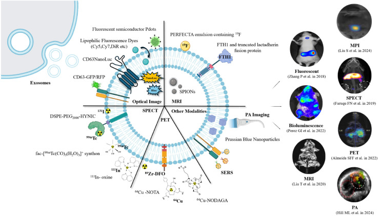
Exosomes-nanoscale extracellular vesicles secreted by various cell types-play a crucial role in intercellular communication by delivering biologically active molecules, such as proteins, nucleic acids, and lipids. Due to their intrinsic biocompatibility, targeting capabilities, and stability, exosomes have emerged as promising vehicles for diagnostics and therapeutics in a wide range of diseases, including cancer, and neurodegenerative, cardiovascular, and autoimmune disorders. The ability to monitor exosome biodistribution and dynamics in vivo is pivotal to promoting their clinical translation. This review provides a comprehensive overview of the current visualization techniques employed for in vivo exosome imaging: optical imaging, magnetic resonance imaging (MRI), single-photon emission computed tomography (SPECT), positron emission tomography (PET), and emerging modalities, such as photoacoustic imaging, ultrasound, and Raman-based approaches. The advantages, limitations, and representative applications of each imaging modality are critically discussed, with emphasis on labeling strategies that enhance tracking sensitivity and specificity. Optical imaging offers high sensitivity, but is limited by shallow tissue penetration, whereas MRI provides excellent spatial resolution, but suffers from low molecular sensitivity. Radionuclide-based imaging, such as PET and SPECT, enables highly sensitive, quantitative tracking, but presents challenges regarding radiotracer stability and safety. Emerging multimodal platforms and labeling innovations are highlighted for their potential to overcome current limitations. Future research directions include the development of non-invasive, highly sensitive, and clinically translatable imaging systems, as well as standardized protocols to ensure reproducibility. Advances in exosome imaging technologies will be instrumental to unlock the full diagnostic and therapeutic potential of exosomebased platforms in precision medicine. [BMB Reports 2025; 58(8): 340-349].
期刊介绍:
The BMB Reports (BMB Rep, established in 1968) is published at the end of every month by Korean Society for Biochemistry and Molecular Biology. Copyright is reserved by the Society. The journal publishes short articles and mini reviews. We expect that the BMB Reports will deliver the new scientific findings and knowledge to our readers in fast and timely manner.
 求助内容:
求助内容: 应助结果提醒方式:


