{"title":"Endotoxaemia in childhood-onset systemic lupus erythematosus induces low-density granulocytes and extracellular traps of innate immune cells.","authors":"Wilasinee Saisorn, Pornpimol Phuengmaung, Chanunya Santiworakul, Kent Doi, Pornpimol Rianthavorn, Asada Leelahavanichkul","doi":"10.1136/lupus-2025-001663","DOIUrl":null,"url":null,"abstract":"<p><strong>Background: </strong>Endotoxaemia without infection in lupus is mentioned with the inconclusive clinical importance.</p><p><strong>Methods: </strong>With endotoxaemia and lupus activity (Systemic Lupus Erythematosus Disease Activity Index 2000 score), 46 patients with childhood-onset lupus were categorised into active lupus with endotoxaemia (n=14), inactive lupus with endotoxaemia (n=10), active lupus without endotoxaemia (n=10) and inactive lupus without endotoxaemia (n=12). The routine parameters (serum creatinine, urine sediments, proteinuria, complement, haematological aspects and histological activity index) were analysed with lupus activity and other parameters.</p><p><strong>Results: </strong>Serum cytokines (tumour necrosis factor (TNF)-α, interleukin (IL)-6, IL-8 and IL-10), serum citrullinated histone H3, cell-free DNA and bacterial-free DNA were not different among groups. The extracellular traps (ETs) in the peripheral blood mononuclear cell (PBMC) fraction, measured by immunofluorescence of myeloperoxidase (MPO) and neutrophil elastase (NE), were elevated in endotoxaemia regardless of lupus disease activity. Interestingly, low-density granulocytes (LDGs), the neutrophils in the PBMC fraction after gradient separation, were elevated in active lupus regardless of endotoxaemia but higher in the patients with positive endotoxaemia. Because endotoxaemia might be derived from the gut, the blood microbiome was measured, and the Burkholderia group was the representative bacteria in active lupus with endotoxaemia. The incubation of LPS or bacterial-free DNA with neutrophils from the healthy control altered these regular-density neutrophils to LDGs.</p><p><strong>Conclusion: </strong>Endotoxaemia presented in both active and inactive lupus (possibly correlated with some bacterial groups in the gut) that caused ETs in the PBMC fraction and LDGs. However, elevated LDGs were most prominent in endotoxaemia with active lupus.</p>","PeriodicalId":18126,"journal":{"name":"Lupus Science & Medicine","volume":"12 2","pages":""},"PeriodicalIF":3.5000,"publicationDate":"2025-07-31","publicationTypes":"Journal Article","fieldsOfStudy":null,"isOpenAccess":false,"openAccessPdf":"https://www.ncbi.nlm.nih.gov/pmc/articles/PMC12314931/pdf/","citationCount":"0","resultStr":null,"platform":"Semanticscholar","paperid":null,"PeriodicalName":"Lupus Science & Medicine","FirstCategoryId":"3","ListUrlMain":"https://doi.org/10.1136/lupus-2025-001663","RegionNum":2,"RegionCategory":"医学","ArticlePicture":[],"TitleCN":null,"AbstractTextCN":null,"PMCID":null,"EPubDate":"","PubModel":"","JCR":"Q1","JCRName":"RHEUMATOLOGY","Score":null,"Total":0}
引用次数: 0
Abstract
Background: Endotoxaemia without infection in lupus is mentioned with the inconclusive clinical importance.
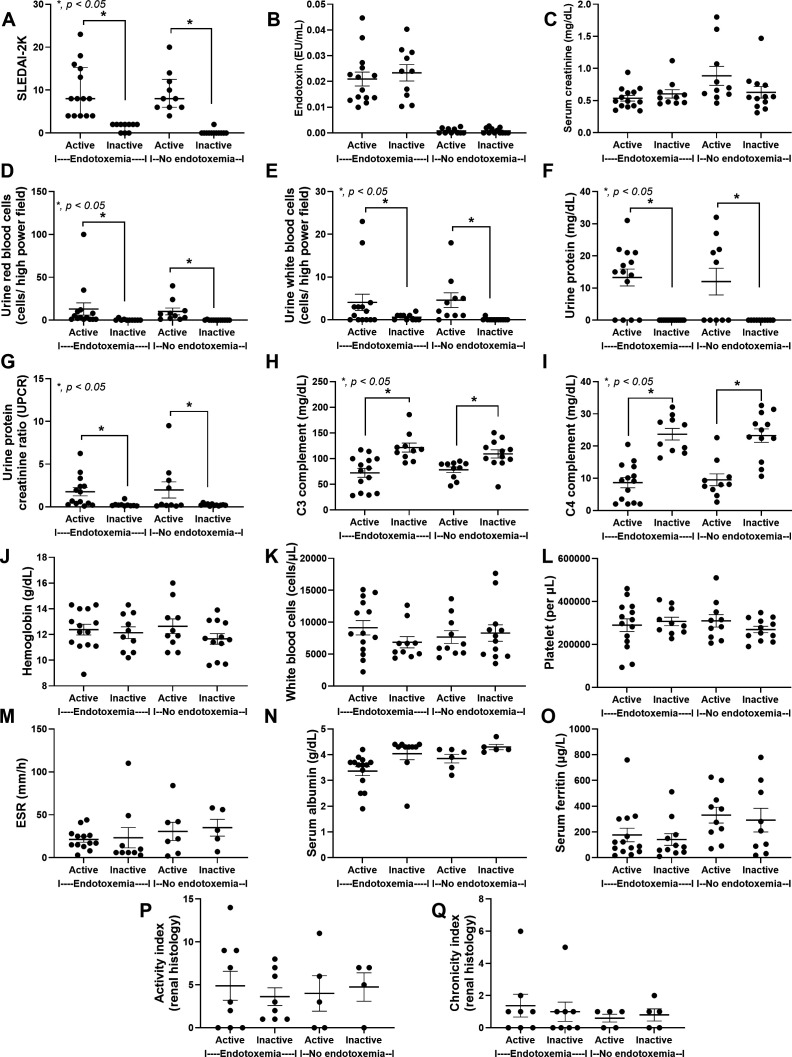
Methods: With endotoxaemia and lupus activity (Systemic Lupus Erythematosus Disease Activity Index 2000 score), 46 patients with childhood-onset lupus were categorised into active lupus with endotoxaemia (n=14), inactive lupus with endotoxaemia (n=10), active lupus without endotoxaemia (n=10) and inactive lupus without endotoxaemia (n=12). The routine parameters (serum creatinine, urine sediments, proteinuria, complement, haematological aspects and histological activity index) were analysed with lupus activity and other parameters.
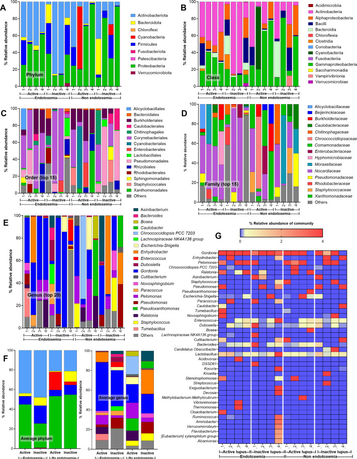
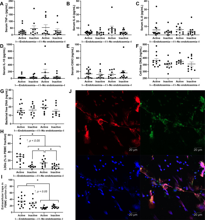
Results: Serum cytokines (tumour necrosis factor (TNF)-α, interleukin (IL)-6, IL-8 and IL-10), serum citrullinated histone H3, cell-free DNA and bacterial-free DNA were not different among groups. The extracellular traps (ETs) in the peripheral blood mononuclear cell (PBMC) fraction, measured by immunofluorescence of myeloperoxidase (MPO) and neutrophil elastase (NE), were elevated in endotoxaemia regardless of lupus disease activity. Interestingly, low-density granulocytes (LDGs), the neutrophils in the PBMC fraction after gradient separation, were elevated in active lupus regardless of endotoxaemia but higher in the patients with positive endotoxaemia. Because endotoxaemia might be derived from the gut, the blood microbiome was measured, and the Burkholderia group was the representative bacteria in active lupus with endotoxaemia. The incubation of LPS or bacterial-free DNA with neutrophils from the healthy control altered these regular-density neutrophils to LDGs.
Conclusion: Endotoxaemia presented in both active and inactive lupus (possibly correlated with some bacterial groups in the gut) that caused ETs in the PBMC fraction and LDGs. However, elevated LDGs were most prominent in endotoxaemia with active lupus.
期刊介绍:
Lupus Science & Medicine is a global, peer reviewed, open access online journal that provides a central point for publication of basic, clinical, translational, and epidemiological studies of all aspects of lupus and related diseases. It is the first lupus-specific open access journal in the world and was developed in response to the need for a barrier-free forum for publication of groundbreaking studies in lupus. The journal publishes research on lupus from fields including, but not limited to: rheumatology, dermatology, nephrology, immunology, pediatrics, cardiology, hepatology, pulmonology, obstetrics and gynecology, and psychiatry.
 求助内容:
求助内容: 应助结果提醒方式:


