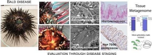
Bald disease in a natural population of the purple sea urchin Paracentrotus lividus of the Mediterranean Sea: From spines to tissues
IF 2.4
3区 生物学
Q1 ZOOLOGY
引用次数: 0
Abstract
Recently, unusual mortality outbreaks have been reported in the echinoderm populations over broad geographic regions. The present work used different diagnostic approaches to unravel the Bald Sea Urchin Disease (BSUD) causes in a natural population of Paracentrotus lividus from the Gulf of Naples sampled in 2021. Symptomatic individuals displayed the typical signs such as test discoloration and ulceration, loss of spines and pedicellariae and visceral hyperpigmentation. Scanning electron microscopy of diseased individuals (stage 2 and stage 3) revealed a bare exoskeleton with multiplying bacteria penetrating the damaged test, and histopathology revealed inflammatory lesions and phagocytosis only in stages 2 and 3, with Gram negative and positive bacteria at stage 3. Metagenomics revealed an increase in DNA virus and Proteobacteria during disease progression. Microbial community analyses failed to reveal a single putative pathogen associated with symptoms, but microbiome analysis showed higher diversity in asymptomatic individuals compared to the symptomatic. Different Vibrio spp. belonging to the Splendidus clade were also isolated, with V. crassostreae as the most represented in advanced stages of disease. We cannot confirm that the observed microorganisms were associated with tissue damage and their contribution to the disease outcome remains unclear as they could be just opportunistic in the lesions. This preliminary study on a wild population highlights the importance of morphological analysis (histopathology and SEM) coupled with microbiome and metagenome analyses in sea urchin disease investigations. Moreover, we suggest also performing transmission electron microscopy (TEM), experimental challenges and in situ hybridization methods (ISH) to provide morphological evidence of potential infective agents. Future studies should also include histopathology of the test.
地中海紫海胆(Paracentrotus lividus)自然种群的秃顶病:从棘到组织。
最近,在广泛地理区域的棘皮动物种群中报告了不寻常的死亡暴发。目前的工作使用不同的诊断方法来解开2021年采样的那不勒斯湾自然种群的秃头海胆病(BSUD)原因。有症状的个体表现出典型的体征,如试验变色和溃疡,脊柱和马蒂的损失和内脏色素沉着。病变个体(第2期和第3期)的扫描电镜显示裸露的外骨骼,大量细菌穿透损伤试验,组织病理学显示炎性病变和吞噬作用仅在第2期和第3期,革兰氏阴性和阳性细菌在第3期。宏基因组学显示,在疾病进展过程中,DNA病毒和变形杆菌增加。微生物群落分析未能揭示与症状相关的单一假定病原体,但与无症状个体相比,无症状个体的微生物群落多样性更高。还分离到了不同弧菌属,属脾弧菌分支,其中在疾病晚期以长弧菌最具代表性。我们不能确认观察到的微生物与组织损伤有关,它们对疾病结果的贡献尚不清楚,因为它们可能只是病变中的机会性微生物。这项对野生种群的初步研究强调了形态学分析(组织病理学和扫描电镜)以及微生物组和宏基因组在海胆疾病调查中的重要性。此外,我们还建议使用透射电子显微镜(TEM)、实验挑战和原位杂交方法(ISH)来提供潜在感染因子的形态学证据。未来的研究还应包括该测试的组织病理学。
本文章由计算机程序翻译,如有差异,请以英文原文为准。
求助全文
约1分钟内获得全文
求助全文
来源期刊
CiteScore
6.10
自引率
5.90%
发文量
94
审稿时长
1 months
期刊介绍:
The Journal of Invertebrate Pathology presents original research articles and notes on the induction and pathogenesis of diseases of invertebrates, including the suppression of diseases in beneficial species, and the use of diseases in controlling undesirable species. In addition, the journal publishes the results of physiological, morphological, genetic, immunological and ecological studies as related to the etiologic agents of diseases of invertebrates.
The Journal of Invertebrate Pathology is the adopted journal of the Society for Invertebrate Pathology, and is available to SIP members at a special reduced price.
 求助内容:
求助内容: 应助结果提醒方式:


