{"title":"Differentiating between adrenocortical carcinoma and pheochromocytoma by a CT-based radiomics model: a multicenter retrospective study.","authors":"Yinyao Chao, Hongzhang Zhu, Wenyi Yang, Haohua Yao, Nan Ma, Xianda Chen, Jing Zhao, Huali Ma, Zhenhua Liu, Hui Han, Zhuowei Liu, Kai Yao, Yiyao Li, Peng Wu, Jingtong Zhang, Bin Li, Shengjie Guo","doi":"10.1186/s12880-025-01842-7","DOIUrl":null,"url":null,"abstract":"<p><strong>Background: </strong>Adrenocortical carcinoma (ACC), a rare and highly malignant adrenal gland tumor, exhibits computed tomography (CT) characteristics that resemble those of the less malignant pheochromocytoma (PHEO). While biochemical evaluation is widely accepted for differentiating between ACC and PHEO, non-functioning tumors remain a diagnostic challenge. The similarity in CT imaging and atypical hormone levels can lead to suboptimal accuracy in diagnosis, leading to inappropriate clinical interventions. This study aims to differentiate between large (≥ 4 cm) ACC and PHEO with radiomics features based on contrast-enhanced CT.</p><p><strong>Methods: </strong>In this retrospective study, 158 patients who received pathological diagnoses of ACC or PHEO between January 2011 and September 2023 were enrolled from three institutions. Radiomics features were extracted from different phases of contrast-enhanced CT and then selected by a two-step procedure. The radiomics model was developed in a cohort of 109 patients from Institution 1, then the model performance was evaluated in the external test cohort of 49 patients from Institutions 2 and 3. The area under the receiver operating characteristic curve (AUC) of the radiomics model was compared with two radiologists using the DeLong test. Hormone testing results were collected to determine the presence of excess cortisol or catecholamines. SHapley Additive exPlanations (SHAP) was used to improve the interpretability of the radiomics model.</p><p><strong>Results: </strong>We developed and evaluated a radiomics model consisting of ten selected CT-based radiomics features. In the external test cohort, the proposed radiomics model achieved high accuracy (86%), specificity (88%), and sensitivity (81%) in differentiating between ACC and PHEO and outperformed 2 radiologists (AUC 0.920 vs. 0.786, 0.629). This radiomics model showed strong capabilities in differentiating biochemically negative ACC and PHEO (with an accuracy of 80%). Moreover, its performance remained consistent even when cortisol and catecholamine levels were simultaneously elevated. Furthermore, SHAP provided quantitative explanations for the radiomics model and visualized the diagnostic process.</p><p><strong>Conclusions: </strong>The interpretable CT-based radiomics model outperforms radiologists in differentiating between ACC and PHEO, especially when hormone testing results are atypical.</p>","PeriodicalId":9020,"journal":{"name":"BMC Medical Imaging","volume":"25 1","pages":"310"},"PeriodicalIF":3.2000,"publicationDate":"2025-08-01","publicationTypes":"Journal Article","fieldsOfStudy":null,"isOpenAccess":false,"openAccessPdf":"https://www.ncbi.nlm.nih.gov/pmc/articles/PMC12317479/pdf/","citationCount":"0","resultStr":null,"platform":"Semanticscholar","paperid":null,"PeriodicalName":"BMC Medical Imaging","FirstCategoryId":"3","ListUrlMain":"https://doi.org/10.1186/s12880-025-01842-7","RegionNum":3,"RegionCategory":"医学","ArticlePicture":[],"TitleCN":null,"AbstractTextCN":null,"PMCID":null,"EPubDate":"","PubModel":"","JCR":"Q2","JCRName":"RADIOLOGY, NUCLEAR MEDICINE & MEDICAL IMAGING","Score":null,"Total":0}
引用次数: 0
Abstract
Background: Adrenocortical carcinoma (ACC), a rare and highly malignant adrenal gland tumor, exhibits computed tomography (CT) characteristics that resemble those of the less malignant pheochromocytoma (PHEO). While biochemical evaluation is widely accepted for differentiating between ACC and PHEO, non-functioning tumors remain a diagnostic challenge. The similarity in CT imaging and atypical hormone levels can lead to suboptimal accuracy in diagnosis, leading to inappropriate clinical interventions. This study aims to differentiate between large (≥ 4 cm) ACC and PHEO with radiomics features based on contrast-enhanced CT.
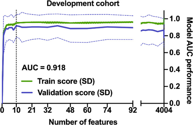
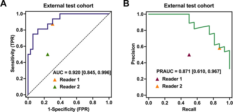
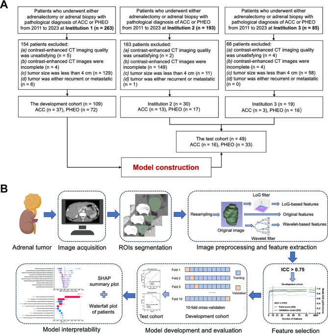
Methods: In this retrospective study, 158 patients who received pathological diagnoses of ACC or PHEO between January 2011 and September 2023 were enrolled from three institutions. Radiomics features were extracted from different phases of contrast-enhanced CT and then selected by a two-step procedure. The radiomics model was developed in a cohort of 109 patients from Institution 1, then the model performance was evaluated in the external test cohort of 49 patients from Institutions 2 and 3. The area under the receiver operating characteristic curve (AUC) of the radiomics model was compared with two radiologists using the DeLong test. Hormone testing results were collected to determine the presence of excess cortisol or catecholamines. SHapley Additive exPlanations (SHAP) was used to improve the interpretability of the radiomics model.
Results: We developed and evaluated a radiomics model consisting of ten selected CT-based radiomics features. In the external test cohort, the proposed radiomics model achieved high accuracy (86%), specificity (88%), and sensitivity (81%) in differentiating between ACC and PHEO and outperformed 2 radiologists (AUC 0.920 vs. 0.786, 0.629). This radiomics model showed strong capabilities in differentiating biochemically negative ACC and PHEO (with an accuracy of 80%). Moreover, its performance remained consistent even when cortisol and catecholamine levels were simultaneously elevated. Furthermore, SHAP provided quantitative explanations for the radiomics model and visualized the diagnostic process.
Conclusions: The interpretable CT-based radiomics model outperforms radiologists in differentiating between ACC and PHEO, especially when hormone testing results are atypical.
期刊介绍:
BMC Medical Imaging is an open access journal publishing original peer-reviewed research articles in the development, evaluation, and use of imaging techniques and image processing tools to diagnose and manage disease.

 求助内容:
求助内容: 应助结果提醒方式:


