Sharjeel Usmani, Khulood Al Riyami, Anjali Jain, Asiya Al Busaidi, Paul Dumasig, Vipin V Jayakrishnan, Subhash Kheruka, Najeeb Ahmed
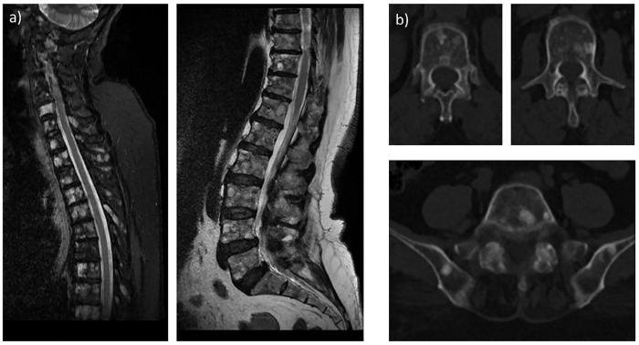
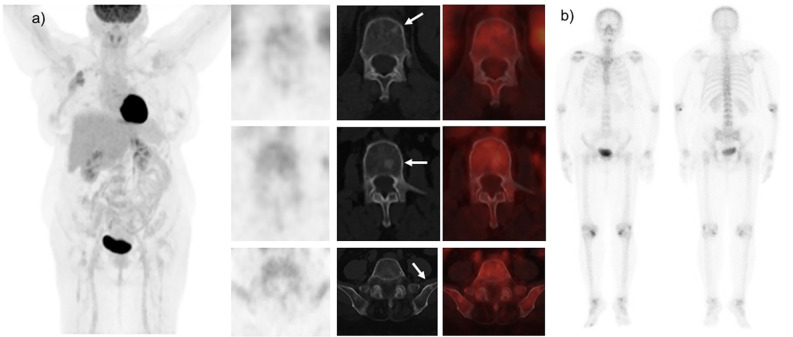
{"title":"Unveiling the Diagnostic Mystery: <sup>18</sup>F-FDG PET and Bone Scan Negative in Bone Metastases of Lobular Breast Cancer: A Case Report.","authors":"Sharjeel Usmani, Khulood Al Riyami, Anjali Jain, Asiya Al Busaidi, Paul Dumasig, Vipin V Jayakrishnan, Subhash Kheruka, Najeeb Ahmed","doi":"10.4274/mirt.galenos.2025.75875","DOIUrl":null,"url":null,"abstract":"<p><p>Identifying osseous metastases by imaging is essential and may be challenging in patients with lobular breast cancer. We present a case of a 66-year-old woman with lobular breast cancer who underwent <sup>18</sup>F- fluorodeoxyglucose positron emission tomography/computed tomography (FDG PET/CT) for staging purposes. <sup>18</sup>F-FDG PET/CT reveals minimal FDG uptake in the primary tumor cells. Prominent sclerotic lesions with low FDG avidity are seen in the spinal and pelvic bones. The subsequent Tc-99m methylene diphosphonate bone scan is unremarkable. The magnetic resonance imaging (MRI) reveals bone metastases. MRI may be beneficial in invasive lobular carcinoma. MRI facilitates improved metastatic evaluation, especially in bone-only and bone-predominant metastatic malignancies, when assessment with <sup>18</sup>F-FDG PET/CT may be difficult and constrained.</p>","PeriodicalId":44681,"journal":{"name":"Molecular Imaging and Radionuclide Therapy","volume":" ","pages":"239-241"},"PeriodicalIF":1.1000,"publicationDate":"2025-10-08","publicationTypes":"Journal Article","fieldsOfStudy":null,"isOpenAccess":false,"openAccessPdf":"https://www.ncbi.nlm.nih.gov/pmc/articles/PMC12505183/pdf/","citationCount":"0","resultStr":null,"platform":"Semanticscholar","paperid":null,"PeriodicalName":"Molecular Imaging and Radionuclide Therapy","FirstCategoryId":"1085","ListUrlMain":"https://doi.org/10.4274/mirt.galenos.2025.75875","RegionNum":0,"RegionCategory":null,"ArticlePicture":[],"TitleCN":null,"AbstractTextCN":null,"PMCID":null,"EPubDate":"2025/8/1 0:00:00","PubModel":"Epub","JCR":"Q4","JCRName":"RADIOLOGY, NUCLEAR MEDICINE & MEDICAL IMAGING","Score":null,"Total":0}
引用次数: 0
Abstract
Identifying osseous metastases by imaging is essential and may be challenging in patients with lobular breast cancer. We present a case of a 66-year-old woman with lobular breast cancer who underwent 18F- fluorodeoxyglucose positron emission tomography/computed tomography (FDG PET/CT) for staging purposes. 18F-FDG PET/CT reveals minimal FDG uptake in the primary tumor cells. Prominent sclerotic lesions with low FDG avidity are seen in the spinal and pelvic bones. The subsequent Tc-99m methylene diphosphonate bone scan is unremarkable. The magnetic resonance imaging (MRI) reveals bone metastases. MRI may be beneficial in invasive lobular carcinoma. MRI facilitates improved metastatic evaluation, especially in bone-only and bone-predominant metastatic malignancies, when assessment with 18F-FDG PET/CT may be difficult and constrained.
期刊介绍:
Molecular Imaging and Radionuclide Therapy (Mol Imaging Radionucl Ther, MIRT) is publishes original research articles, invited reviews, editorials, short communications, letters, consensus statements, guidelines and case reports with a literature review on the topic, in the field of molecular imaging, multimodality imaging, nuclear medicine, radionuclide therapy, radiopharmacy, medical physics, dosimetry and radiobiology.

 求助内容:
求助内容: 应助结果提醒方式:


