Analysis of Imaging Findings in a Patient with Squamous Cell Carcinoma of the External Auditory Canal Metastatic to the Dura with Trigeminal Nerve Involvement.
IF 1.1
Q4 RADIOLOGY, NUCLEAR MEDICINE & MEDICAL IMAGING
Molecular Imaging and Radionuclide Therapy
Pub Date : 2025-10-08
Epub Date: 2025-08-01
DOI:10.4274/mirt.galenos.2025.26937
引用次数: 0
Abstract
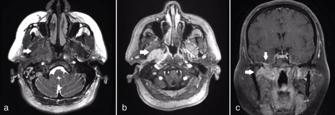
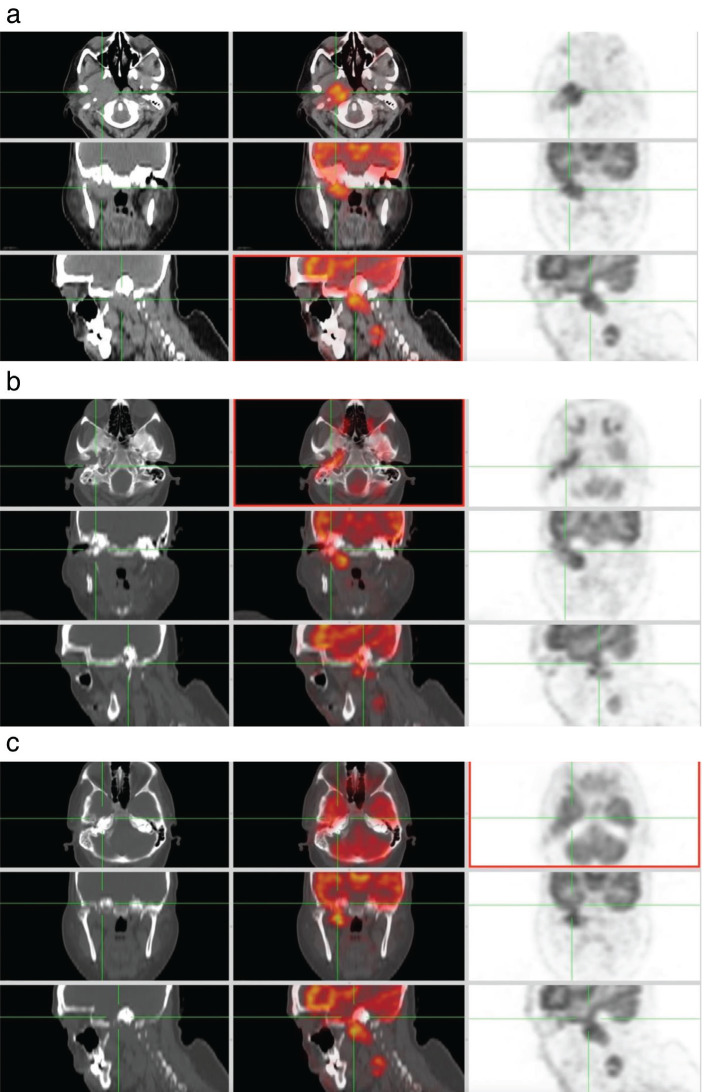
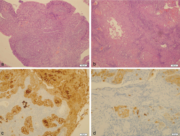
We report the case of a 56-year-old female recently diagnosed with well-differentiated squamous cell carcinoma of the external auditory canal. The patient underwent an 18F-fluorodeoxyglucose positron emission tomography/computed tomography for staging assessment. This examination revealed intense uptake in the right ear canals, tympanic cavity, eustachian canal, parapharyngeal area, and infratemporal fossa. Notably, we identified intracranial dural metastasis, which represents an uncommon site for metastatic spread in general.
外耳道鳞状细胞癌转移至硬脑膜并累及三叉神经1例影像学分析。
我们报告一个56岁的女性最近被诊断为外耳道高分化鳞状细胞癌的情况。患者接受了18f氟脱氧葡萄糖正电子发射断层扫描/计算机断层扫描进行分期评估。检查发现在右耳道、鼓室、耳咽管、咽旁区和颞下窝有强烈的摄取。值得注意的是,我们发现了颅内硬脑膜转移,这是一种不常见的转移扩散部位。
本文章由计算机程序翻译,如有差异,请以英文原文为准。
求助全文
约1分钟内获得全文
求助全文
来源期刊

Molecular Imaging and Radionuclide Therapy
RADIOLOGY, NUCLEAR MEDICINE & MEDICAL IMAGING-
CiteScore
1.30
自引率
0.00%
发文量
50
期刊介绍:
Molecular Imaging and Radionuclide Therapy (Mol Imaging Radionucl Ther, MIRT) is publishes original research articles, invited reviews, editorials, short communications, letters, consensus statements, guidelines and case reports with a literature review on the topic, in the field of molecular imaging, multimodality imaging, nuclear medicine, radionuclide therapy, radiopharmacy, medical physics, dosimetry and radiobiology.
 求助内容:
求助内容: 应助结果提醒方式:


