{"title":"Evaluation of TTF-1, Napsin A, p40, and p63 in the Subtyping of Non-Small Cell Lung Carcinoma: A Cross-Sectional Study from India.","authors":"Surbhi Patel, Deepa Sowkur Anandarama Adiga","doi":"10.30699/ijp.2025.2044252.3371","DOIUrl":null,"url":null,"abstract":"<p><strong>Background & objective: </strong>Subtyping non-small cell lung carcinoma (NSCLC) into adenocarcinoma (ADC) and squamous cell carcinoma (SCC) is crucial for selecting appropriate molecular tests, as driver mutations are often subtype-specific. This study aimed to evaluate the utility of TTF-1, Napsin A, p40, and p63 immunohistochemical (IHC) markers in subtyping NSCLC on small biopsies, with the goal of identifying a minimal marker panel.</p><p><strong>Methods: </strong>This retrospective, cross-sectional study was conducted at Kasturba Medical College, Mangalore, from January 2014 to December 2020. All NSCLC cases diagnosed during the study period were included. Immunohistochemical expressions of TTF-1, Napsin A, p40, and p63 were evaluated and correlated with morphological findings.</p><p><strong>Results: </strong>Ninety-five NSCLC cases were included: adenocarcinoma (n = 35), squamous cell carcinoma (n = 57), and NSCLC-not otherwise specified (NOS) (n = 2). IHC reclassification based on marker expression resulted in six ADC cases retyped as SCC and eight SCC cases retyped as ADC. TTF-1 and Napsin A expression were significantly associated with adenocarcinoma (<i>p</i> < 0.001), while p40 and p63 expression were significantly associated with SCC (<i>p</i> < 0.001).</p><p><strong>Conclusion: </strong>IHC is essential in overcoming the diagnostic limitations of small biopsy specimens, especially in morphologically heterogeneous tumors. A minimal panel comprising TTF-1 and p40 is sufficient for accurate subtyping of NSCLC and can help preserve tissue for downstream molecular testing.</p>","PeriodicalId":38900,"journal":{"name":"Iranian Journal of Pathology","volume":"20 3","pages":"297-306"},"PeriodicalIF":0.0000,"publicationDate":"2025-01-01","publicationTypes":"Journal Article","fieldsOfStudy":null,"isOpenAccess":false,"openAccessPdf":"https://www.ncbi.nlm.nih.gov/pmc/articles/PMC12308192/pdf/","citationCount":"0","resultStr":null,"platform":"Semanticscholar","paperid":null,"PeriodicalName":"Iranian Journal of Pathology","FirstCategoryId":"1085","ListUrlMain":"https://doi.org/10.30699/ijp.2025.2044252.3371","RegionNum":0,"RegionCategory":null,"ArticlePicture":[],"TitleCN":null,"AbstractTextCN":null,"PMCID":null,"EPubDate":"2025/7/1 0:00:00","PubModel":"Epub","JCR":"Q3","JCRName":"Medicine","Score":null,"Total":0}
引用次数: 0
Abstract
Background & objective: Subtyping non-small cell lung carcinoma (NSCLC) into adenocarcinoma (ADC) and squamous cell carcinoma (SCC) is crucial for selecting appropriate molecular tests, as driver mutations are often subtype-specific. This study aimed to evaluate the utility of TTF-1, Napsin A, p40, and p63 immunohistochemical (IHC) markers in subtyping NSCLC on small biopsies, with the goal of identifying a minimal marker panel.
Methods: This retrospective, cross-sectional study was conducted at Kasturba Medical College, Mangalore, from January 2014 to December 2020. All NSCLC cases diagnosed during the study period were included. Immunohistochemical expressions of TTF-1, Napsin A, p40, and p63 were evaluated and correlated with morphological findings.
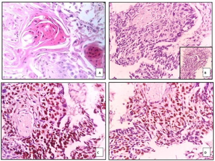
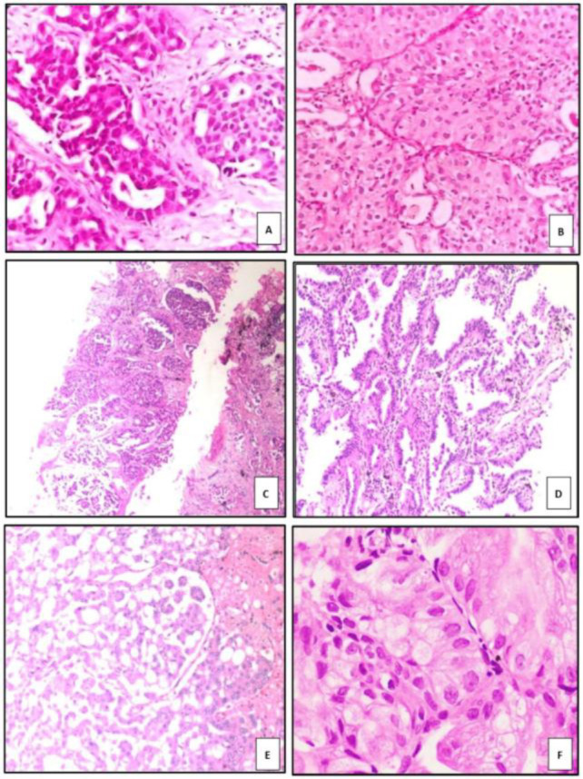
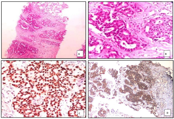
Results: Ninety-five NSCLC cases were included: adenocarcinoma (n = 35), squamous cell carcinoma (n = 57), and NSCLC-not otherwise specified (NOS) (n = 2). IHC reclassification based on marker expression resulted in six ADC cases retyped as SCC and eight SCC cases retyped as ADC. TTF-1 and Napsin A expression were significantly associated with adenocarcinoma (p < 0.001), while p40 and p63 expression were significantly associated with SCC (p < 0.001).
Conclusion: IHC is essential in overcoming the diagnostic limitations of small biopsy specimens, especially in morphologically heterogeneous tumors. A minimal panel comprising TTF-1 and p40 is sufficient for accurate subtyping of NSCLC and can help preserve tissue for downstream molecular testing.

 求助内容:
求助内容: 应助结果提醒方式:


