Fahime Norozi, Mehdi Allahbakhshian, Nader Vazifeshiran, Zahra Hasanpour, Mohsen Hamidpour
{"title":"Evaluation of HOTAIRM1, miR-196b, and HOXA9 as Oncogenic Markers in Patients with Acute Myeloblastic Leukemia.","authors":"Fahime Norozi, Mehdi Allahbakhshian, Nader Vazifeshiran, Zahra Hasanpour, Mohsen Hamidpour","doi":"10.30699/ijp.2025.2030358.3309","DOIUrl":null,"url":null,"abstract":"<p><strong>Background & objective: </strong>miR-196b, HOXA9, GFI1, and PIM1 are key factors involved in cellular signaling pathways that contribute to the pathogenesis of malignancies, including acute myeloblastic leukemia (AML). Given their critical roles in AML progression, the present study aimed to investigate the gene expression levels of HOTAIRM1, miR-196b, HOXA9, GFI1, and PIM1 in AML patients compared to healthy controls.</p><p><strong>Methods: </strong>A total of 30 AML patients and 10 healthy volunteers were enrolled in this study. Peripheral blood and bone marrow mononuclear cells were isolated using Ficoll-Paque density gradient centrifugation. Gene expression levels of HOTAIRM1, miR-196b, HOXA9, GFI1, and PIM1 were assessed using real-time quantitative PCR (RQ-PCR). Statistical analyses were performed using Student's t-test, one-way ANOVA, and Pearson correlation tests.</p><p><strong>Results: </strong>The expression levels of HOTAIRM1, miR-196b, HOXA9, and GFI1 were significantly elevated in AML patients compared to healthy controls. Furthermore, t-test analysis revealed that the expressions of HOTAIRM1, HOXA9, and GFI1 significantly differed between AML-M3 and non-M3 AML subtypes.</p><p><strong>Conclusion: </strong>These findings suggest that the investigated markers, particularly HOTAIRM1, HOXA9, and GFI1, may serve as potential clinical biomarkers for monitoring AML progression and could be valuable targets for early detection or therapeutic intervention.</p>","PeriodicalId":38900,"journal":{"name":"Iranian Journal of Pathology","volume":"20 3","pages":"307-315"},"PeriodicalIF":0.0000,"publicationDate":"2025-01-01","publicationTypes":"Journal Article","fieldsOfStudy":null,"isOpenAccess":false,"openAccessPdf":"https://www.ncbi.nlm.nih.gov/pmc/articles/PMC12308185/pdf/","citationCount":"0","resultStr":null,"platform":"Semanticscholar","paperid":null,"PeriodicalName":"Iranian Journal of Pathology","FirstCategoryId":"1085","ListUrlMain":"https://doi.org/10.30699/ijp.2025.2030358.3309","RegionNum":0,"RegionCategory":null,"ArticlePicture":[],"TitleCN":null,"AbstractTextCN":null,"PMCID":null,"EPubDate":"2025/7/1 0:00:00","PubModel":"Epub","JCR":"Q3","JCRName":"Medicine","Score":null,"Total":0}
引用次数: 0
Abstract
Background & objective: miR-196b, HOXA9, GFI1, and PIM1 are key factors involved in cellular signaling pathways that contribute to the pathogenesis of malignancies, including acute myeloblastic leukemia (AML). Given their critical roles in AML progression, the present study aimed to investigate the gene expression levels of HOTAIRM1, miR-196b, HOXA9, GFI1, and PIM1 in AML patients compared to healthy controls.
Methods: A total of 30 AML patients and 10 healthy volunteers were enrolled in this study. Peripheral blood and bone marrow mononuclear cells were isolated using Ficoll-Paque density gradient centrifugation. Gene expression levels of HOTAIRM1, miR-196b, HOXA9, GFI1, and PIM1 were assessed using real-time quantitative PCR (RQ-PCR). Statistical analyses were performed using Student's t-test, one-way ANOVA, and Pearson correlation tests.
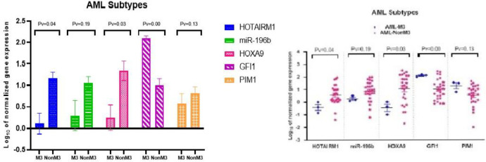
Results: The expression levels of HOTAIRM1, miR-196b, HOXA9, and GFI1 were significantly elevated in AML patients compared to healthy controls. Furthermore, t-test analysis revealed that the expressions of HOTAIRM1, HOXA9, and GFI1 significantly differed between AML-M3 and non-M3 AML subtypes.
Conclusion: These findings suggest that the investigated markers, particularly HOTAIRM1, HOXA9, and GFI1, may serve as potential clinical biomarkers for monitoring AML progression and could be valuable targets for early detection or therapeutic intervention.


 求助内容:
求助内容: 应助结果提醒方式:


