Adam R Collins, Gerard M O'Connor, Darragh A Ryan, Molly Parmeter, Sean Dinneen, Georgina Gethin
{"title":"Wound Bed Temperature has Potential to Indicate Infection Status: A Cross-Sectional Study.","authors":"Adam R Collins, Gerard M O'Connor, Darragh A Ryan, Molly Parmeter, Sean Dinneen, Georgina Gethin","doi":"10.1111/wrr.70072","DOIUrl":null,"url":null,"abstract":"<p><p>This study investigates the potential of wound bed temperature, measured using an IR camera, to aid in the clinical assessment of chronic wounds. The study captured thermal images from 267 patients with chronic wounds (diabetic foot ulcers, pressure ulcers, venous leg ulcers and arterial ulcers) with corresponding photographic images and clinical data. Temperature measurements were extracted from thermal images, focusing on both the centre of the wound and the surrounding periwound skin. Statistical analyses were employed to evaluate the relationship between wound temperature distribution and clinical diagnosis. The results showed a strong correlation between wound centre temperature and the average temperature across the entire wound (R<sup>2</sup> = 0.977). This indicates that a single-point measurement is representative of the entire wound, simplifying wound temperature assessment. A fair correlation was found between the temperature difference between the wound and periwound and the clinician's assessment of infection status (Pearson coefficient = 0.32). The study concludes that thermal imaging holds promise as a supplementary tool for clinicians in assessing chronic wound status, especially in cases where infection is unclear. It is a low-cost, non-contact, and easy-to-use technique.</p>","PeriodicalId":23864,"journal":{"name":"Wound Repair and Regeneration","volume":"33 4","pages":"e70072"},"PeriodicalIF":3.4000,"publicationDate":"2025-07-01","publicationTypes":"Journal Article","fieldsOfStudy":null,"isOpenAccess":false,"openAccessPdf":"https://www.ncbi.nlm.nih.gov/pmc/articles/PMC12315627/pdf/","citationCount":"0","resultStr":null,"platform":"Semanticscholar","paperid":null,"PeriodicalName":"Wound Repair and Regeneration","FirstCategoryId":"3","ListUrlMain":"https://doi.org/10.1111/wrr.70072","RegionNum":3,"RegionCategory":"医学","ArticlePicture":[],"TitleCN":null,"AbstractTextCN":null,"PMCID":null,"EPubDate":"","PubModel":"","JCR":"Q2","JCRName":"CELL BIOLOGY","Score":null,"Total":0}
引用次数: 0
Abstract
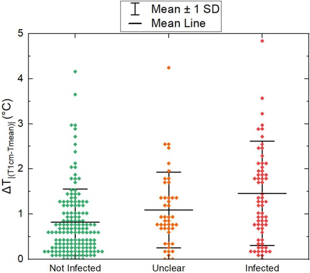
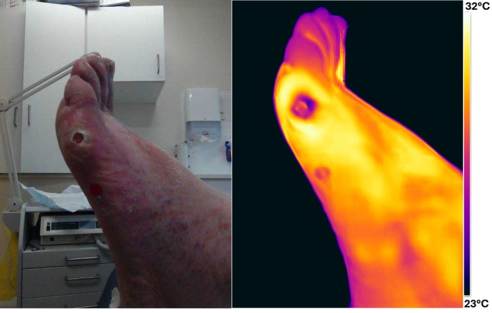
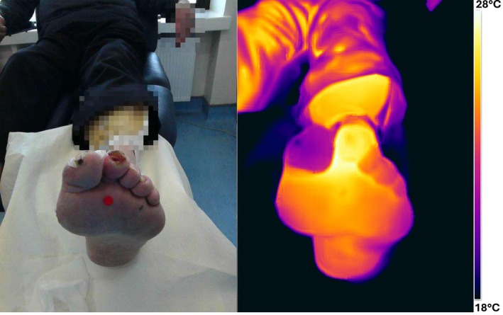
This study investigates the potential of wound bed temperature, measured using an IR camera, to aid in the clinical assessment of chronic wounds. The study captured thermal images from 267 patients with chronic wounds (diabetic foot ulcers, pressure ulcers, venous leg ulcers and arterial ulcers) with corresponding photographic images and clinical data. Temperature measurements were extracted from thermal images, focusing on both the centre of the wound and the surrounding periwound skin. Statistical analyses were employed to evaluate the relationship between wound temperature distribution and clinical diagnosis. The results showed a strong correlation between wound centre temperature and the average temperature across the entire wound (R2 = 0.977). This indicates that a single-point measurement is representative of the entire wound, simplifying wound temperature assessment. A fair correlation was found between the temperature difference between the wound and periwound and the clinician's assessment of infection status (Pearson coefficient = 0.32). The study concludes that thermal imaging holds promise as a supplementary tool for clinicians in assessing chronic wound status, especially in cases where infection is unclear. It is a low-cost, non-contact, and easy-to-use technique.
期刊介绍:
Wound Repair and Regeneration provides extensive international coverage of cellular and molecular biology, connective tissue, and biological mediator studies in the field of tissue repair and regeneration and serves a diverse audience of surgeons, plastic surgeons, dermatologists, biochemists, cell biologists, and others.
Wound Repair and Regeneration is the official journal of The Wound Healing Society, The European Tissue Repair Society, The Japanese Society for Wound Healing, and The Australian Wound Management Association.
 求助内容:
求助内容: 应助结果提醒方式:


