{"title":"Differentiation of Cerebral Cystic Echinococcosis (CCE) from Coenurosis Using Morphometric and Molecular Methods.","authors":"Fattaneh Mikaeili, Sharif Maraghi, Eshrat Beigom Kia, Fatemeh Sadat Sadjjadi, Mehdi Karamian, Seyed Shahram Shekarforoush, Majid Fasihi Harandi, Seyed Mahmoud Sadjjadi","doi":"10.18502/ijpa.v20i2.19023","DOIUrl":null,"url":null,"abstract":"<p><p>Cerebral cystic echinococcosis (CCE) and coenurosis are zoonotic diseases caused by the larval stages of <i>Echinococcus granulosus sensu lato (s.l.)</i> and <i>Taenia</i> spp., respectively. Due to the similarity between the symptoms and clinical samples of CCE and cerebral coenurosis, especially in cases with no protoscoleces, the diagnostic methods for the differentiation of CCE from cerebral coenurosis are crucial, especially in countries where both diseases are endemic. To compare CCE and coenurosis, morphometric indices of protoscoleces and molecular methods were used in the present study.</p><p><strong>Methods: </strong>In this regard, four isolates of human cerebral echinococcal cysts, three isolates of <i>Coenurus cerebralis</i> from sheep, and one non-cerebral <i>Coenurus</i> from sheep muscles were evaluated. The isolated specimens have been collected from Shiraz, Ahvaz, Tehran and Kerman from before 2000 to 2022. The molecular characterization was carried out using the partial NADH dehydrogenase1 (nad1) gene. Phylogenetic analysis was performed using the maximum likelihood method.</p><p><strong>Results: </strong>In fertile cysts, the total size of the large and small hooks of <i>Coenurus</i> was larger than cerebral echinococcal cyst. These parameters demonstrated significant morphological differences between the <i>C. cerebralis</i> and the cerebral echinococcal cyst. Molecular methods identified the cerebral echinococcal cysts as <i>E. canadensis</i> (G6) genotype. One <i>C. cerebralis</i> and the non-cerebral <i>Coenurus</i> were identified as <i>Taenia multiceps</i> and <i>T. multiceps gaigeri</i>, respectively.</p><p><strong>Conclusion: </strong>Morphometric indices are significantly different between protoscoleces of <i>C. cerebralis</i> and cerebral echinococcal cysts. Hence, they could be used for differential diagnosis of the fertile cysts of these cestodes. However, in cases with no protoscoleces, molecular methods are essential for the differentiation of CCE from cerebral coenurosis, especially in regions where both diseases are prevalent and endemic.</p>","PeriodicalId":14669,"journal":{"name":"Iranian Journal of Parasitology","volume":"20 2","pages":"193-202"},"PeriodicalIF":0.9000,"publicationDate":"2025-04-01","publicationTypes":"Journal Article","fieldsOfStudy":null,"isOpenAccess":false,"openAccessPdf":"https://www.ncbi.nlm.nih.gov/pmc/articles/PMC12307782/pdf/","citationCount":"0","resultStr":null,"platform":"Semanticscholar","paperid":null,"PeriodicalName":"Iranian Journal of Parasitology","FirstCategoryId":"3","ListUrlMain":"https://doi.org/10.18502/ijpa.v20i2.19023","RegionNum":4,"RegionCategory":"医学","ArticlePicture":[],"TitleCN":null,"AbstractTextCN":null,"PMCID":null,"EPubDate":"","PubModel":"","JCR":"Q4","JCRName":"PARASITOLOGY","Score":null,"Total":0}
引用次数: 0
Abstract
Cerebral cystic echinococcosis (CCE) and coenurosis are zoonotic diseases caused by the larval stages of Echinococcus granulosus sensu lato (s.l.) and Taenia spp., respectively. Due to the similarity between the symptoms and clinical samples of CCE and cerebral coenurosis, especially in cases with no protoscoleces, the diagnostic methods for the differentiation of CCE from cerebral coenurosis are crucial, especially in countries where both diseases are endemic. To compare CCE and coenurosis, morphometric indices of protoscoleces and molecular methods were used in the present study.
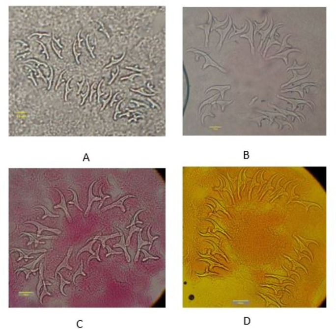
Methods: In this regard, four isolates of human cerebral echinococcal cysts, three isolates of Coenurus cerebralis from sheep, and one non-cerebral Coenurus from sheep muscles were evaluated. The isolated specimens have been collected from Shiraz, Ahvaz, Tehran and Kerman from before 2000 to 2022. The molecular characterization was carried out using the partial NADH dehydrogenase1 (nad1) gene. Phylogenetic analysis was performed using the maximum likelihood method.
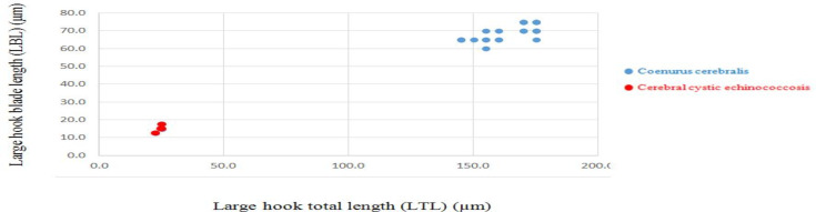
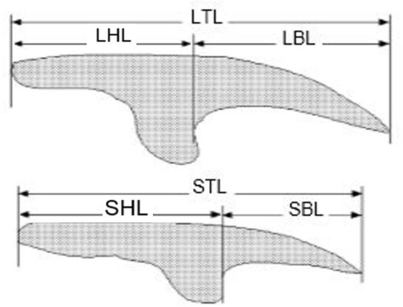
Results: In fertile cysts, the total size of the large and small hooks of Coenurus was larger than cerebral echinococcal cyst. These parameters demonstrated significant morphological differences between the C. cerebralis and the cerebral echinococcal cyst. Molecular methods identified the cerebral echinococcal cysts as E. canadensis (G6) genotype. One C. cerebralis and the non-cerebral Coenurus were identified as Taenia multiceps and T. multiceps gaigeri, respectively.
Conclusion: Morphometric indices are significantly different between protoscoleces of C. cerebralis and cerebral echinococcal cysts. Hence, they could be used for differential diagnosis of the fertile cysts of these cestodes. However, in cases with no protoscoleces, molecular methods are essential for the differentiation of CCE from cerebral coenurosis, especially in regions where both diseases are prevalent and endemic.
期刊介绍:
Iranian Journal of Parasitology (IJP) is the official publication of Iranian Society of Parasitology (ISP) launched in 2006. The society was inaugurated in 1994 and pursues the improvement of the knowledge on the parasites and parasitic diseases, exchange of scientific knowledge with foreign societies, publicity activities, and consultation on the parasitic diseases, and intimate relationship among society members.
The main aims of the Journal are: contribution to the field of Parasitology, including all aspects of parasites and parasitic diseases (medical and veterinary) and related fields such as Entomology which may be submitted by scientists from Iran and all over the world.
 求助内容:
求助内容: 应助结果提醒方式:


