A cell fate mapping simulation laboratory to increase undergraduate students' understanding of early developmental processes in frog, zebrafish, and tunicate embryos.
{"title":"A cell fate mapping simulation laboratory to increase undergraduate students' understanding of early developmental processes in frog, zebrafish, and tunicate embryos.","authors":"Ritu Sarpal, Ashley E E Bruce, Isaac Skromne","doi":"10.1128/jmbe.00104-25","DOIUrl":null,"url":null,"abstract":"<p><p>Fate mapping is an essential technique in developmental biology that allows researchers to track the future identity or \"fate\" of embryonic cells in an organism. However, the experimental procedure for constructing fate maps is tedious, time-consuming, and technically challenging, making it difficult to incorporate as an undergraduate lab experience. Here, we describe a hands-on undergraduate laboratory activity that allows students to generate and examine model organisms' fate maps, employing a free, user-friendly web-based app, FatemapApp (http://fatemapapp.com/). Students used the app to construct the fate maps for the 32-cell stage <i>Xenopus laevis</i> frog embryo, the gastrula stage <i>Danio rerio</i> zebrafish embryo, and the 76-cell stage <i>Holocynthia roretzi</i> tunicate embryo. Individual analysis of the maps allows students to identify the potential of cells to contribute to one or multiple tissues and their probability of moving and mixing with the neighboring cells. Subsequently, cross-species comparative analysis allows students to infer tissue organization across chordate and vertebrate embryos that may be evolutionarily conserved. Surveys showed that the students found this activity engaging and valuable, reporting a deeper understanding of the rationale, methodology, and outcomes underlying the construction of fate maps. Furthermore, students reported increased comprehension of embryonic development and its processes.</p>","PeriodicalId":46416,"journal":{"name":"Journal of Microbiology & Biology Education","volume":" ","pages":"e0010425"},"PeriodicalIF":1.5000,"publicationDate":"2025-08-21","publicationTypes":"Journal Article","fieldsOfStudy":null,"isOpenAccess":false,"openAccessPdf":"https://www.ncbi.nlm.nih.gov/pmc/articles/PMC12369337/pdf/","citationCount":"0","resultStr":null,"platform":"Semanticscholar","paperid":null,"PeriodicalName":"Journal of Microbiology & Biology Education","FirstCategoryId":"1085","ListUrlMain":"https://doi.org/10.1128/jmbe.00104-25","RegionNum":0,"RegionCategory":null,"ArticlePicture":[],"TitleCN":null,"AbstractTextCN":null,"PMCID":null,"EPubDate":"2025/7/31 0:00:00","PubModel":"Epub","JCR":"Q2","JCRName":"EDUCATION, SCIENTIFIC DISCIPLINES","Score":null,"Total":0}
引用次数: 0
Abstract
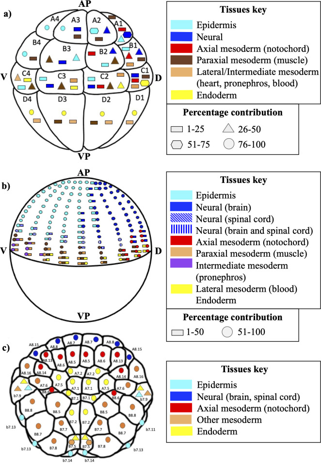
Fate mapping is an essential technique in developmental biology that allows researchers to track the future identity or "fate" of embryonic cells in an organism. However, the experimental procedure for constructing fate maps is tedious, time-consuming, and technically challenging, making it difficult to incorporate as an undergraduate lab experience. Here, we describe a hands-on undergraduate laboratory activity that allows students to generate and examine model organisms' fate maps, employing a free, user-friendly web-based app, FatemapApp (http://fatemapapp.com/). Students used the app to construct the fate maps for the 32-cell stage Xenopus laevis frog embryo, the gastrula stage Danio rerio zebrafish embryo, and the 76-cell stage Holocynthia roretzi tunicate embryo. Individual analysis of the maps allows students to identify the potential of cells to contribute to one or multiple tissues and their probability of moving and mixing with the neighboring cells. Subsequently, cross-species comparative analysis allows students to infer tissue organization across chordate and vertebrate embryos that may be evolutionarily conserved. Surveys showed that the students found this activity engaging and valuable, reporting a deeper understanding of the rationale, methodology, and outcomes underlying the construction of fate maps. Furthermore, students reported increased comprehension of embryonic development and its processes.

 求助内容:
求助内容: 应助结果提醒方式:


