Chest ultrasound vs. Radiograph for pneumothorax diagnosis performed by emergency healthcare workers in the emergency department: a systematic review and meta-analysis.
{"title":"Chest ultrasound vs. Radiograph for pneumothorax diagnosis performed by emergency healthcare workers in the emergency department: a systematic review and meta-analysis.","authors":"Jean-Baptiste Bouillon-Minois, Coline Burlet, Resa E Lewiss, Reza Bagheri, Christophe Perrier, Jeannot Schmidt, Frédéric Dutheil","doi":"10.1186/s13089-025-00441-5","DOIUrl":null,"url":null,"abstract":"<p><strong>Background: </strong>The efficacy of bedside chest ultrasonography for the detection and diagnosis of pneumothorax is under debate. We aimed to compare Emergency Healthcare Workers performed chest ultrasonography with chest X-ray in the detection and diagnosis of pneumothorax in the emergency department.</p><p><strong>Methods: </strong>We queried PubMed, Cochrane, ScienceDirect, Web of Science and ClinicalTrials.gov databases from 2000 through January 2024. We included all studies (both retrospective and prospective) that compared the diagnostic performance of chest ultrasonography with chest radiography, using chest computed tomography as the gold standard. Participants are patients consulting in the emergency department and physician that performed the chest ultrasound was an Emergency Healthcare Workers. Studies reporting the sensitivity and specificity for both chest ultrasonography and chest X-ray met inclusion criteria. We applied a random effects meta-analysis methodology. We then performed a meta-regression analysis to search for influencing variables such as technical parameters of echograph, patients and pneumothorax.</p><p><strong>Main results: </strong>15 studies totaling 3,171 patients were analyzed. 71% of patients were male with a mean age of 40.2 years. The mean prevalence of pneumothorax was 27.6% (95 CI 20.9 to 34.3). Chest ultrasonography had higher sensitivity (79.4%, 68.2 to 90.7) compared to chest X-ray (48.1%, 36.8 to 59.4), and a greater negative predictive value (chest ultrasonography = 94.3%, 91.2 to 97.3, and chest X-ray = 87.9%, 84.1 to 91.6). There was no statistical difference in specificity between the two modalities: chest ultrasonography 99.5%, 99 to 100 and chest X-ray 99.8%, 99.4 to 100) or in positive predictive value (chest ultrasonography 94.2%, 90.5 to 97.9 vs chest X-ray 96.7%,92 to 100). Characteristics of echograph or pneumothorax and patients sociodemographic did not influence results.</p><p><strong>Conclusion: </strong>In this systematic review and meta-analysis, chest ultrasonography performed by Emergency Healthcare Workers, had greater sensitivity and negative predictive value than chest radiography for the diagnosis of pneumothorax in emergency department patients.</p>","PeriodicalId":36911,"journal":{"name":"Ultrasound Journal","volume":"17 1","pages":"37"},"PeriodicalIF":2.9000,"publicationDate":"2025-07-31","publicationTypes":"Journal Article","fieldsOfStudy":null,"isOpenAccess":false,"openAccessPdf":"https://www.ncbi.nlm.nih.gov/pmc/articles/PMC12314293/pdf/","citationCount":"0","resultStr":null,"platform":"Semanticscholar","paperid":null,"PeriodicalName":"Ultrasound Journal","FirstCategoryId":"1085","ListUrlMain":"https://doi.org/10.1186/s13089-025-00441-5","RegionNum":0,"RegionCategory":null,"ArticlePicture":[],"TitleCN":null,"AbstractTextCN":null,"PMCID":null,"EPubDate":"","PubModel":"","JCR":"Q2","JCRName":"Medicine","Score":null,"Total":0}
引用次数: 0
Abstract
Background: The efficacy of bedside chest ultrasonography for the detection and diagnosis of pneumothorax is under debate. We aimed to compare Emergency Healthcare Workers performed chest ultrasonography with chest X-ray in the detection and diagnosis of pneumothorax in the emergency department.
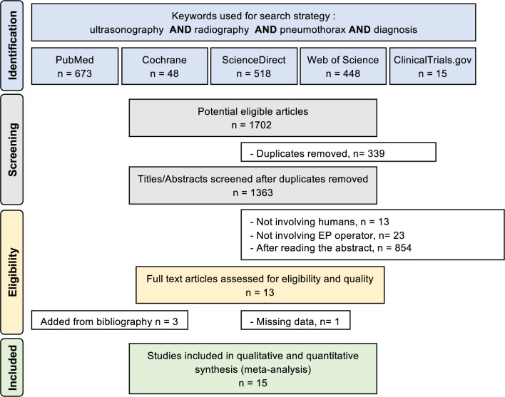
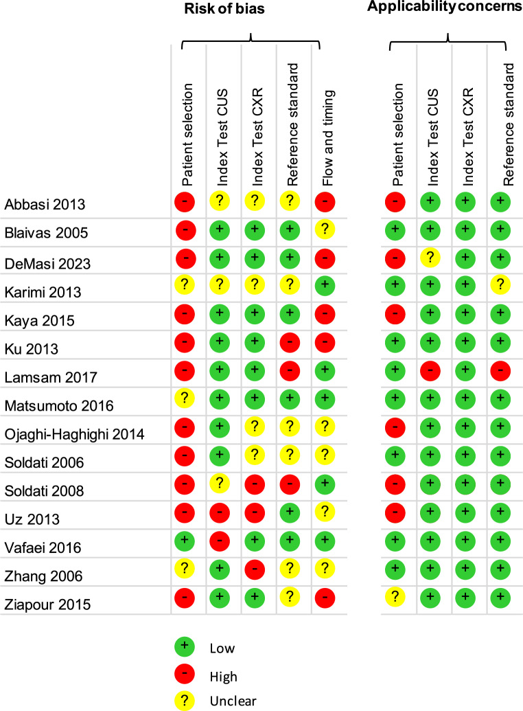
Methods: We queried PubMed, Cochrane, ScienceDirect, Web of Science and ClinicalTrials.gov databases from 2000 through January 2024. We included all studies (both retrospective and prospective) that compared the diagnostic performance of chest ultrasonography with chest radiography, using chest computed tomography as the gold standard. Participants are patients consulting in the emergency department and physician that performed the chest ultrasound was an Emergency Healthcare Workers. Studies reporting the sensitivity and specificity for both chest ultrasonography and chest X-ray met inclusion criteria. We applied a random effects meta-analysis methodology. We then performed a meta-regression analysis to search for influencing variables such as technical parameters of echograph, patients and pneumothorax.
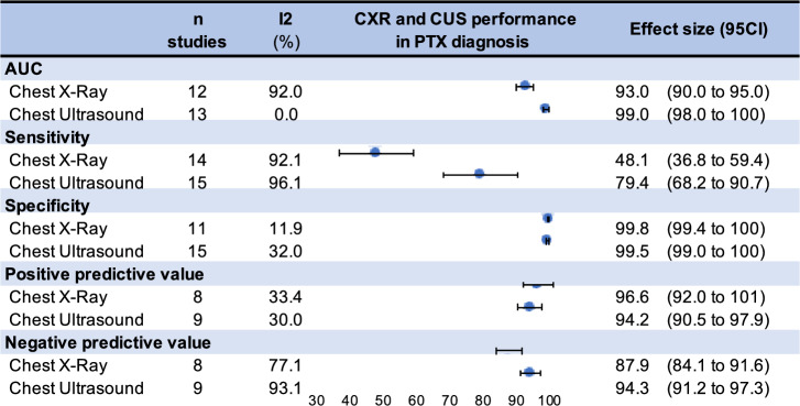
Main results: 15 studies totaling 3,171 patients were analyzed. 71% of patients were male with a mean age of 40.2 years. The mean prevalence of pneumothorax was 27.6% (95 CI 20.9 to 34.3). Chest ultrasonography had higher sensitivity (79.4%, 68.2 to 90.7) compared to chest X-ray (48.1%, 36.8 to 59.4), and a greater negative predictive value (chest ultrasonography = 94.3%, 91.2 to 97.3, and chest X-ray = 87.9%, 84.1 to 91.6). There was no statistical difference in specificity between the two modalities: chest ultrasonography 99.5%, 99 to 100 and chest X-ray 99.8%, 99.4 to 100) or in positive predictive value (chest ultrasonography 94.2%, 90.5 to 97.9 vs chest X-ray 96.7%,92 to 100). Characteristics of echograph or pneumothorax and patients sociodemographic did not influence results.
Conclusion: In this systematic review and meta-analysis, chest ultrasonography performed by Emergency Healthcare Workers, had greater sensitivity and negative predictive value than chest radiography for the diagnosis of pneumothorax in emergency department patients.

 求助内容:
求助内容: 应助结果提醒方式:


