{"title":"A comparative analysis of manual glenoid version measurement using two-dimensional and three-dimensional computed tomography imaging techniques.","authors":"Maxwell S Renna, Ashley I Simpson","doi":"10.5397/cise.2025.00318","DOIUrl":null,"url":null,"abstract":"<p><strong>Background: </strong>Accurate measurement of glenoid version is essential for optimal implant positioning in shoulder arthroplasty. This study compared the accuracy and reliability of unformatted two-dimensional computed tomography (2D-CT), formatted 2D-CT, and three-dimensional CT (3D-CT) reconstructions in measuring glenoid version.</p><p><strong>Methods: </strong>Shoulder CT scans for arthroplasty were analyzed retrospectively. Glenoid version was measured at the estimated glenoid midpoint using unformatted and formatted 2D-CT in the scapular plane. Measurements from 3D-CT reconstructions using the Corrected Friedman Method were used as the control. Inter- and intra-observer reliability was calculated as well as minimally detectable difference.</p><p><strong>Results: </strong>Sixty-five CT scans were analyzed (mean age, 61.7 years). Mean glenoid version was -3.48° (standard deviation [SD], 8.7°) on unformatted 2D-CT, -3.27° (SD, 8.15°) on formatted 2D-CT, and -4.25° (SD, 7.92°) on 3D-CT. Although no significant difference in mean values was observed (analysis of variance, P=0.245), formatted 2D-CT measurements were within 6° of 3D-CT in 95.4% of cases versus 83.1% for unformatted 2D-CT (P=0.023). Directional error occurred in 27.7% of unformatted scans and 16.9% of formatted scans. Inter-observer reliability was highest for 3D-CT (intraclass correlation coefficient [ICC]=0.83; 95% CI, 0.74-0.89), and intra-observer agreement was strongest for 3D-CT (ICC=0.91; 95% CI, 0.86-0.94), followed by formatted 2D-CT (ICC=0.83; 95% CI, 0.73-0.89) and unformatted 2D-CT (ICC=0.77; 95% CI, 0.65-0.85).</p><p><strong>Conclusions: </strong>3D-CT reconstructions are widely considered the most accurate and reproducible method for glenoid version assessment, supported by multiple comparative imaging studies. Formatted 2D-CT provides a reliable alternative when 3D-CT is unavailable, significantly outperforming unformatted 2D-CT in both agreement with the 3D reference and intra- and inter-observer reliability. Level of evidence: IV.</p>","PeriodicalId":33981,"journal":{"name":"Clinics in Shoulder and Elbow","volume":" ","pages":""},"PeriodicalIF":1.7000,"publicationDate":"2025-07-31","publicationTypes":"Journal Article","fieldsOfStudy":null,"isOpenAccess":false,"openAccessPdf":"https://www.ncbi.nlm.nih.gov/pmc/articles/PMC12415454/pdf/","citationCount":"0","resultStr":null,"platform":"Semanticscholar","paperid":null,"PeriodicalName":"Clinics in Shoulder and Elbow","FirstCategoryId":"1085","ListUrlMain":"https://doi.org/10.5397/cise.2025.00318","RegionNum":0,"RegionCategory":null,"ArticlePicture":[],"TitleCN":null,"AbstractTextCN":null,"PMCID":null,"EPubDate":"","PubModel":"","JCR":"Q2","JCRName":"ORTHOPEDICS","Score":null,"Total":0}
引用次数: 0
Abstract
Background: Accurate measurement of glenoid version is essential for optimal implant positioning in shoulder arthroplasty. This study compared the accuracy and reliability of unformatted two-dimensional computed tomography (2D-CT), formatted 2D-CT, and three-dimensional CT (3D-CT) reconstructions in measuring glenoid version.
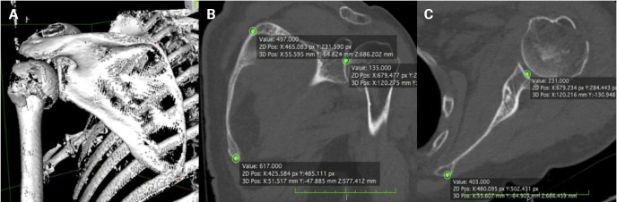
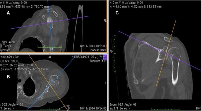
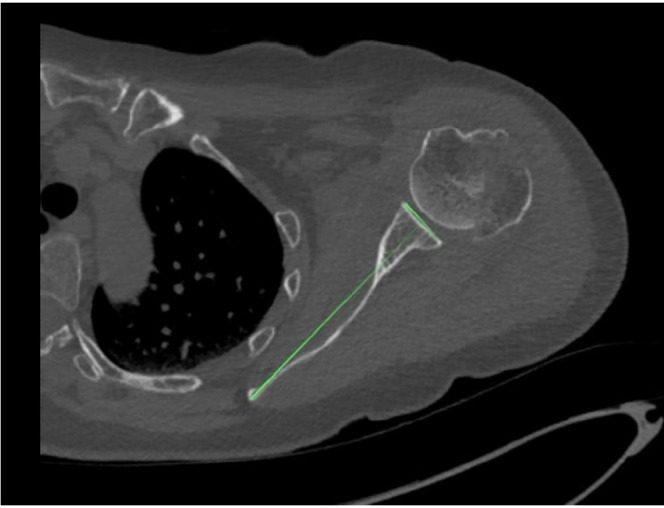
Methods: Shoulder CT scans for arthroplasty were analyzed retrospectively. Glenoid version was measured at the estimated glenoid midpoint using unformatted and formatted 2D-CT in the scapular plane. Measurements from 3D-CT reconstructions using the Corrected Friedman Method were used as the control. Inter- and intra-observer reliability was calculated as well as minimally detectable difference.
Results: Sixty-five CT scans were analyzed (mean age, 61.7 years). Mean glenoid version was -3.48° (standard deviation [SD], 8.7°) on unformatted 2D-CT, -3.27° (SD, 8.15°) on formatted 2D-CT, and -4.25° (SD, 7.92°) on 3D-CT. Although no significant difference in mean values was observed (analysis of variance, P=0.245), formatted 2D-CT measurements were within 6° of 3D-CT in 95.4% of cases versus 83.1% for unformatted 2D-CT (P=0.023). Directional error occurred in 27.7% of unformatted scans and 16.9% of formatted scans. Inter-observer reliability was highest for 3D-CT (intraclass correlation coefficient [ICC]=0.83; 95% CI, 0.74-0.89), and intra-observer agreement was strongest for 3D-CT (ICC=0.91; 95% CI, 0.86-0.94), followed by formatted 2D-CT (ICC=0.83; 95% CI, 0.73-0.89) and unformatted 2D-CT (ICC=0.77; 95% CI, 0.65-0.85).
Conclusions: 3D-CT reconstructions are widely considered the most accurate and reproducible method for glenoid version assessment, supported by multiple comparative imaging studies. Formatted 2D-CT provides a reliable alternative when 3D-CT is unavailable, significantly outperforming unformatted 2D-CT in both agreement with the 3D reference and intra- and inter-observer reliability. Level of evidence: IV.

 求助内容:
求助内容: 应助结果提醒方式:


