Fei Wang, Chuan Huang, Hai-Qing Ma, Xue-Qing Yao, Jun-Jiang Wang, Jie Long
{"title":"Mesentery morphological features on computed tomography for preoperative prediction of tumor invasion and lymph node metastasis in colon cancer.","authors":"Fei Wang, Chuan Huang, Hai-Qing Ma, Xue-Qing Yao, Jun-Jiang Wang, Jie Long","doi":"10.5306/wjco.v16.i7.108095","DOIUrl":null,"url":null,"abstract":"<p><strong>Background: </strong>Accurate identification of tumor invasion depth and lymph node (LN) involvement in patients with colon cancer (CC) is critical for guiding treatment strategies. However, the preoperative prediction of tumor invasion depth and LN metastasis in CC remains challenging. As the intestinal tumor develops, the fat density in the mesentery increases.</p><p><strong>Aim: </strong>To investigate the efficacy of computed tomography (CT) value change in the mesentery contributed by the tumor (CT-T value) for predicting tumor invasion depth and LN metastasis.</p><p><strong>Methods: </strong>Patients, who were diagnosed with CC and underwent surgery, were included and divided into the training and validation cohorts. CT-T values of the mesentery were extracted from the CT images. Cutoff points were determined using the receiver operating characteristic (ROC) curve, and the area under the ROC curve was employed to assess the performance of the CT-T value for tumor invasion depth and LN status prediction.</p><p><strong>Results: </strong>Cutoff values of 11.83 and 17.17 were identified to discriminate T1/2 <i>vs</i> T3/4 and N0 <i>vs</i> N1/2, respectively. With a cutoff CT-T value of 11.83, the total diagnostic accuracy for T stage was 83.1% (81.5% for the training cohort and 86.2% for the validation cohort). With a cutoff CT-T value of 17.17, the total diagnostic accuracy for N stage was 77.3% (75.8% for the training cohort and 80.1% for the validation cohort), which was higher than that of CT-reported LN metastasis.</p><p><strong>Conclusion: </strong>In this study, we explored an efficient method for predicting preoperative T and N stages using the tumor-contributed CT value of the mesentery in CC, which displayed superior predictive accuracy.</p>","PeriodicalId":23802,"journal":{"name":"World journal of clinical oncology","volume":"16 7","pages":"108095"},"PeriodicalIF":3.2000,"publicationDate":"2025-07-24","publicationTypes":"Journal Article","fieldsOfStudy":null,"isOpenAccess":false,"openAccessPdf":"https://www.ncbi.nlm.nih.gov/pmc/articles/PMC12305001/pdf/","citationCount":"0","resultStr":null,"platform":"Semanticscholar","paperid":null,"PeriodicalName":"World journal of clinical oncology","FirstCategoryId":"1085","ListUrlMain":"https://doi.org/10.5306/wjco.v16.i7.108095","RegionNum":0,"RegionCategory":null,"ArticlePicture":[],"TitleCN":null,"AbstractTextCN":null,"PMCID":null,"EPubDate":"","PubModel":"","JCR":"Q3","JCRName":"ONCOLOGY","Score":null,"Total":0}
引用次数: 0
Abstract
Background: Accurate identification of tumor invasion depth and lymph node (LN) involvement in patients with colon cancer (CC) is critical for guiding treatment strategies. However, the preoperative prediction of tumor invasion depth and LN metastasis in CC remains challenging. As the intestinal tumor develops, the fat density in the mesentery increases.
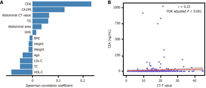
Aim: To investigate the efficacy of computed tomography (CT) value change in the mesentery contributed by the tumor (CT-T value) for predicting tumor invasion depth and LN metastasis.
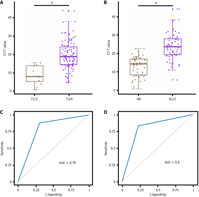
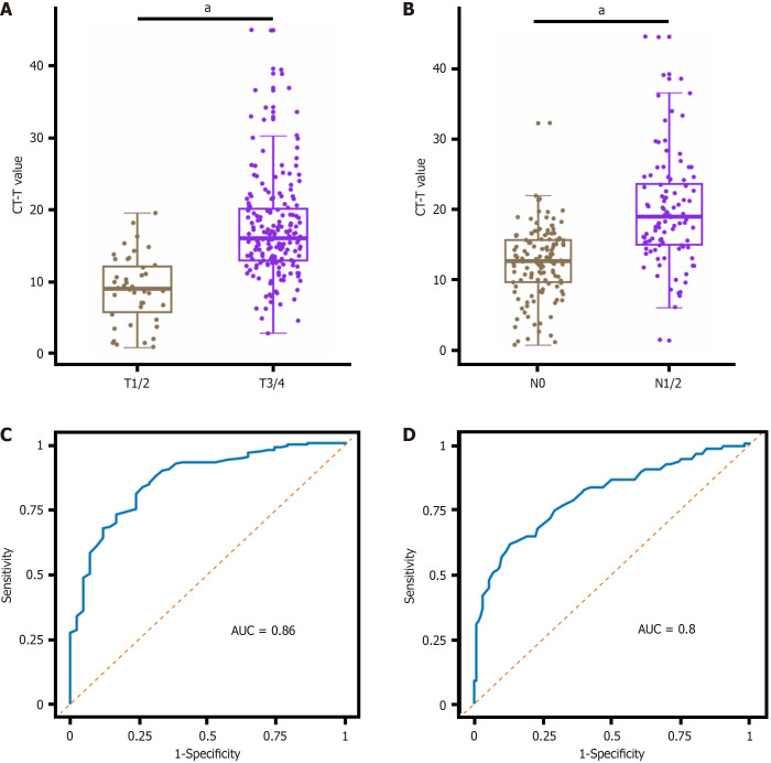
Methods: Patients, who were diagnosed with CC and underwent surgery, were included and divided into the training and validation cohorts. CT-T values of the mesentery were extracted from the CT images. Cutoff points were determined using the receiver operating characteristic (ROC) curve, and the area under the ROC curve was employed to assess the performance of the CT-T value for tumor invasion depth and LN status prediction.
Results: Cutoff values of 11.83 and 17.17 were identified to discriminate T1/2 vs T3/4 and N0 vs N1/2, respectively. With a cutoff CT-T value of 11.83, the total diagnostic accuracy for T stage was 83.1% (81.5% for the training cohort and 86.2% for the validation cohort). With a cutoff CT-T value of 17.17, the total diagnostic accuracy for N stage was 77.3% (75.8% for the training cohort and 80.1% for the validation cohort), which was higher than that of CT-reported LN metastasis.
Conclusion: In this study, we explored an efficient method for predicting preoperative T and N stages using the tumor-contributed CT value of the mesentery in CC, which displayed superior predictive accuracy.
期刊介绍:
The WJCO is a high-quality, peer reviewed, open-access journal. The primary task of WJCO is to rapidly publish high-quality original articles, reviews, editorials, and case reports in the field of oncology. In order to promote productive academic communication, the peer review process for the WJCO is transparent; to this end, all published manuscripts are accompanied by the anonymized reviewers’ comments as well as the authors’ responses. The primary aims of the WJCO are to improve diagnostic, therapeutic and preventive modalities and the skills of clinicians and to guide clinical practice in oncology. Scope: Art of Oncology, Biology of Neoplasia, Breast Cancer, Cancer Prevention and Control, Cancer-Related Complications, Diagnosis in Oncology, Gastrointestinal Cancer, Genetic Testing For Cancer, Gynecologic Cancer, Head and Neck Cancer, Hematologic Malignancy, Lung Cancer, Melanoma, Molecular Oncology, Neurooncology, Palliative and Supportive Care, Pediatric Oncology, Surgical Oncology, Translational Oncology, and Urologic Oncology.

 求助内容:
求助内容: 应助结果提醒方式:


