{"title":"Retinal parameter analysis and diagnostic potentail exploration in familial exudative vitreoretinopathy using ultra-widefield fundus photography.","authors":"Jiayu Li, Shaochi Zhang, Xiaolong Qi, Chanjuan Wang, Wen Zhang, Rui Li, Caihong Sun, Keyan Liu, Xiaolu Li, Wenjuan Zhuang","doi":"10.1186/s40942-025-00716-y","DOIUrl":null,"url":null,"abstract":"<p><strong>Background: </strong>Familial Exudative Vitreoretinopathy (FEVR) is a monogenic disorder causing retinal vascular impairment, often underdiagnosed due to its variable presentation and reliance on invasive methods like fundus fluorescein angiography (FFA). Through the utilization of non-invasive ultra-widefield fundus photography (UWFFP), this research explored both the diagnostic potential of integrated retinal parameters for the detection of FEVR, and their characteristic changes during the early stage progression.</p><p><strong>Methods: </strong>Retinal parameters were systematically extracted and quantified from UWFFP of 114 FEVR patients and 114 matched controls using the EVision AI cloud platform. Comparative statistical analyses were performed to identify significant intergroup differences between FEVR and control cohorts, and assess intra-group variations among FEVR subgroups. Based on parameters that showed significant differences between the FEVR group and the control group and had an impact on the FEVR group, a diagnostic model was constructed. Receiver operating characteristic (ROC) curves were plotted to determine the diagnostic potential of these parameters. In addition, subgroup analysis within the FEVR group was conducted to clarify the relationship between retinal parameters and disease staging.</p><p><strong>Results: </strong>Significant differences were observed in 25 retinal parameters between the FEVR group and the control group, with the horizontal cup-to-disc ratio, vertical cup-to-disc ratio, optic disc-to-macula distance, and vascular density demonstrating potential diagnostic efficacy. Subgroup analysis within the FEVR group revealed that as the disease stage advanced and severity increased, the optic disc and cup diameters decreased, the optic disc-to-macula distance increased, and the vascular fractal dimension and vascular density parameters declined.</p><p><strong>Conclusions: </strong>UWFFP and automated retinal parameter analysis offer promising tools for early FEVR diagnosis, with specific structural and vascular markers providing diagnostic potential. Further large-scale studies are needed to validate these findings and refine diagnostic models.</p>","PeriodicalId":14289,"journal":{"name":"International Journal of Retina and Vitreous","volume":"11 1","pages":"87"},"PeriodicalIF":2.4000,"publicationDate":"2025-07-30","publicationTypes":"Journal Article","fieldsOfStudy":null,"isOpenAccess":false,"openAccessPdf":"https://www.ncbi.nlm.nih.gov/pmc/articles/PMC12312263/pdf/","citationCount":"0","resultStr":null,"platform":"Semanticscholar","paperid":null,"PeriodicalName":"International Journal of Retina and Vitreous","FirstCategoryId":"1085","ListUrlMain":"https://doi.org/10.1186/s40942-025-00716-y","RegionNum":0,"RegionCategory":null,"ArticlePicture":[],"TitleCN":null,"AbstractTextCN":null,"PMCID":null,"EPubDate":"","PubModel":"","JCR":"Q2","JCRName":"OPHTHALMOLOGY","Score":null,"Total":0}
引用次数: 0
Abstract
Background: Familial Exudative Vitreoretinopathy (FEVR) is a monogenic disorder causing retinal vascular impairment, often underdiagnosed due to its variable presentation and reliance on invasive methods like fundus fluorescein angiography (FFA). Through the utilization of non-invasive ultra-widefield fundus photography (UWFFP), this research explored both the diagnostic potential of integrated retinal parameters for the detection of FEVR, and their characteristic changes during the early stage progression.
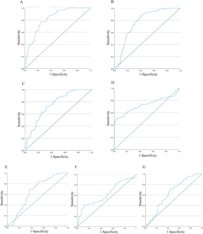
Methods: Retinal parameters were systematically extracted and quantified from UWFFP of 114 FEVR patients and 114 matched controls using the EVision AI cloud platform. Comparative statistical analyses were performed to identify significant intergroup differences between FEVR and control cohorts, and assess intra-group variations among FEVR subgroups. Based on parameters that showed significant differences between the FEVR group and the control group and had an impact on the FEVR group, a diagnostic model was constructed. Receiver operating characteristic (ROC) curves were plotted to determine the diagnostic potential of these parameters. In addition, subgroup analysis within the FEVR group was conducted to clarify the relationship between retinal parameters and disease staging.
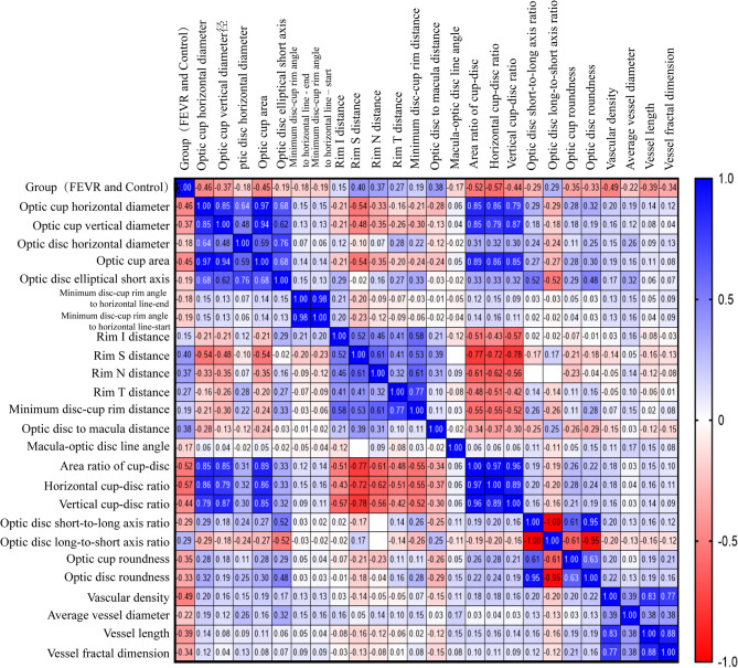
Results: Significant differences were observed in 25 retinal parameters between the FEVR group and the control group, with the horizontal cup-to-disc ratio, vertical cup-to-disc ratio, optic disc-to-macula distance, and vascular density demonstrating potential diagnostic efficacy. Subgroup analysis within the FEVR group revealed that as the disease stage advanced and severity increased, the optic disc and cup diameters decreased, the optic disc-to-macula distance increased, and the vascular fractal dimension and vascular density parameters declined.
Conclusions: UWFFP and automated retinal parameter analysis offer promising tools for early FEVR diagnosis, with specific structural and vascular markers providing diagnostic potential. Further large-scale studies are needed to validate these findings and refine diagnostic models.
期刊介绍:
International Journal of Retina and Vitreous focuses on the ophthalmic subspecialty of vitreoretinal disorders. The journal presents original articles on new approaches to diagnosis, outcomes of clinical trials, innovations in pharmacological therapy and surgical techniques, as well as basic science advances that impact clinical practice. Topical areas include, but are not limited to: -Imaging of the retina, choroid and vitreous -Innovations in optical coherence tomography (OCT) -Small-gauge vitrectomy, retinal detachment, chromovitrectomy -Electroretinography (ERG), microperimetry, other functional tests -Intraocular tumors -Retinal pharmacotherapy & drug delivery -Diabetic retinopathy & other vascular diseases -Age-related macular degeneration (AMD) & other macular entities

 求助内容:
求助内容: 应助结果提醒方式:


