{"title":"Spontaneous Necrosis of Hepatocellular Carcinoma in Viral B Cirrhosis: A Case Report.","authors":"Yasmine Hidous, Abdelwaheb Nakhli, Emna Mrabet, Nesrine Hemdani, Zeineb Benzarti, Bochra Bouchabou, Rym Ennaifer","doi":"10.1159/000546699","DOIUrl":null,"url":null,"abstract":"<p><strong>Introduction: </strong>Hepatocellular carcinoma (HCC) is a leading cause of cancer-related mortality worldwide and its prognosis largely depends on the stage at diagnosis and the feasibility of curative treatments. Spontaneous necrosis of HCC is an extremely rare phenomenon with an unclear pathophysiology. Various mechanisms, including vascular disturbances, immune-mediated response, and recurrent infection, have been considered. This case report presents a rare case of spontaneous necrosis of a small HCC in a patient with hepatitis B-related cirrhosis.</p><p><strong>Case presentation: </strong>A 63-year-old male with compensated hepatitis B-related cirrhosis was under routine surveillance when a suspicious liver lesion was detected on ultrasound. Subsequent contrast-enhanced computed tomography (CT) confirmed the presence of an 18-mm HCC in segment VIII, corresponding to Barcelona Clinical Liver Cancer stage A. Due to limited curative treatment options, transarterial chemoembolization was considered. However, a follow-up CT scan was performed 2 weeks before the procedure unexpectedly revealed complete regression of arterial enhancement, suggestive of spontaneous tumor necrosis. The patient remained asymptomatic, with stable liver function and mild biological inflammatory markers. No evidence of vascular thrombosis or significant systemic inflammation was noted, suggesting localized vascular disturbances or intrinsic tumor factors might have precipitated the necrosis.</p><p><strong>Conclusion: </strong>This case highlights the exceptional occurrence of spontaneous necrosis in a small HCC. While the underlying mechanisms remain speculative, further documentation and research on similar cases may provide insights into HCC pathophysiology and potential implications for future therapeutic strategies.</p>","PeriodicalId":9625,"journal":{"name":"Case Reports in Oncology","volume":"18 1","pages":"1028-1033"},"PeriodicalIF":0.7000,"publicationDate":"2025-06-27","publicationTypes":"Journal Article","fieldsOfStudy":null,"isOpenAccess":false,"openAccessPdf":"https://www.ncbi.nlm.nih.gov/pmc/articles/PMC12310190/pdf/","citationCount":"0","resultStr":null,"platform":"Semanticscholar","paperid":null,"PeriodicalName":"Case Reports in Oncology","FirstCategoryId":"1085","ListUrlMain":"https://doi.org/10.1159/000546699","RegionNum":0,"RegionCategory":null,"ArticlePicture":[],"TitleCN":null,"AbstractTextCN":null,"PMCID":null,"EPubDate":"2025/1/1 0:00:00","PubModel":"eCollection","JCR":"Q4","JCRName":"ONCOLOGY","Score":null,"Total":0}
引用次数: 0
Abstract
Introduction: Hepatocellular carcinoma (HCC) is a leading cause of cancer-related mortality worldwide and its prognosis largely depends on the stage at diagnosis and the feasibility of curative treatments. Spontaneous necrosis of HCC is an extremely rare phenomenon with an unclear pathophysiology. Various mechanisms, including vascular disturbances, immune-mediated response, and recurrent infection, have been considered. This case report presents a rare case of spontaneous necrosis of a small HCC in a patient with hepatitis B-related cirrhosis.
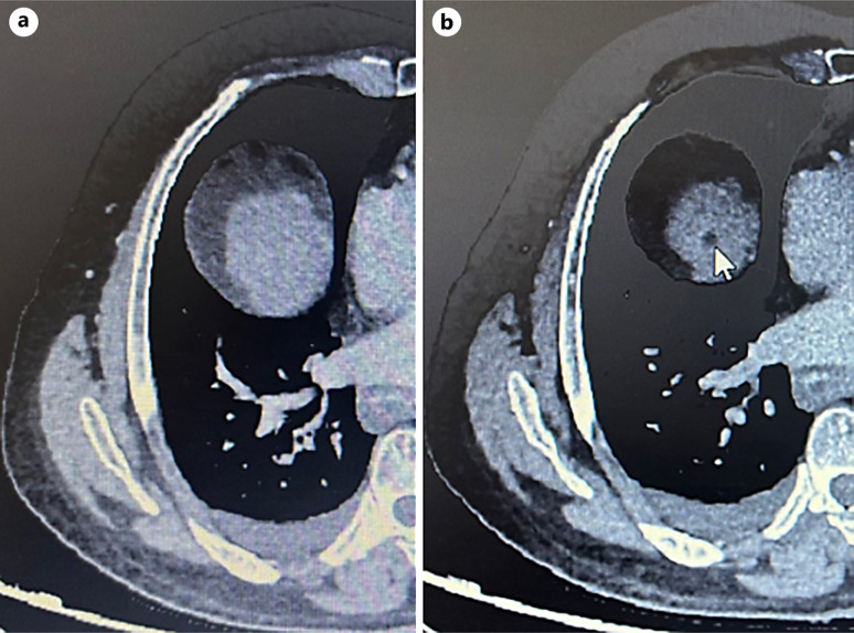
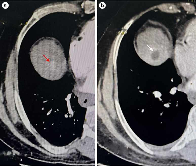
Case presentation: A 63-year-old male with compensated hepatitis B-related cirrhosis was under routine surveillance when a suspicious liver lesion was detected on ultrasound. Subsequent contrast-enhanced computed tomography (CT) confirmed the presence of an 18-mm HCC in segment VIII, corresponding to Barcelona Clinical Liver Cancer stage A. Due to limited curative treatment options, transarterial chemoembolization was considered. However, a follow-up CT scan was performed 2 weeks before the procedure unexpectedly revealed complete regression of arterial enhancement, suggestive of spontaneous tumor necrosis. The patient remained asymptomatic, with stable liver function and mild biological inflammatory markers. No evidence of vascular thrombosis or significant systemic inflammation was noted, suggesting localized vascular disturbances or intrinsic tumor factors might have precipitated the necrosis.
Conclusion: This case highlights the exceptional occurrence of spontaneous necrosis in a small HCC. While the underlying mechanisms remain speculative, further documentation and research on similar cases may provide insights into HCC pathophysiology and potential implications for future therapeutic strategies.

 求助内容:
求助内容: 应助结果提醒方式:


