Jeewoo Choi, Jae-Yong Nam, Min Sung Kim, You Won Choi, Hae Young Choi, Ji Yeon Byun
{"title":"Immunophenotypic and Transcriptomic Analysis of Peripheral Blood Mononuclear Cells in Bullous Pemphigoid.","authors":"Jeewoo Choi, Jae-Yong Nam, Min Sung Kim, You Won Choi, Hae Young Choi, Ji Yeon Byun","doi":"10.5021/ad.25.032","DOIUrl":null,"url":null,"abstract":"<p><strong>Background: </strong>Bullous pemphigoid (BP) is an autoimmune blistering disease driven by autoantibodies against BP180 and BP230. While type 2 inflammation plays a key role, the precise immune cell alterations and transcriptomic changes remain unclear.</p><p><strong>Objective: </strong>To characterize immune cell composition and transcriptomic changes in BP patients using fluorescence-activated cell sorting (FACS)-based immunophenotyping and RNA sequencing.</p><p><strong>Methods: </strong>A case-control study was conducted on 10 newly diagnosed, treatment-naive BP patients and six healthy controls. Disease activity was assessed using the Bullous Pemphigoid Disease Area Index (BPDAI). Peripheral blood mononuclear cells were isolated for FACS analysis to determine immune cell subsets. RNA sequencing was performed to identify differentially expressed genes (DEGs) and enriched pathways. Statistical analyses included t-tests, Mann-Whitney U tests, and correlation analysis.</p><p><strong>Results: </strong>FACS analysis revealed a reduction in CD4⁺ T cells, T helper 2 (Th2), and B cells in BP patients (<i>p</i><0.05), alongside an increase in M2a-like monocytes (<i>p</i><0.001). RNA sequencing identified 262 DEGs, with secretory leukocyte peptidase inhibitor (SLPI) and transmembrane protein 237 being the most significantly upregulated. Proline-serine-threonine phosphatase interacting protein 2 (PSTPIP2) and SAM domain, SH3 domain, and nuclear localization signals 1 (SAMSN1) positively correlated with BPDAI (<i>p</i><0.001). Gene ontology analysis highlighted enrichment in inflammatory responses and neutrophil degranulation pathways.</p><p><strong>Conclusion: </strong>BP patients show distinct immune dysregulation, including decreased CD4⁺ T cells, Th2, and B cells, increased M2a-like monocytes, altered gene expression profiles, and correlations between PSTPIP2, SAMSN1 and disease activity. These findings provide insights into pathogenesis and potential therapeutic targets of BP.</p>","PeriodicalId":94298,"journal":{"name":"Annals of dermatology","volume":"37 4","pages":"191-200"},"PeriodicalIF":1.3000,"publicationDate":"2025-08-01","publicationTypes":"Journal Article","fieldsOfStudy":null,"isOpenAccess":false,"openAccessPdf":"https://www.ncbi.nlm.nih.gov/pmc/articles/PMC12318784/pdf/","citationCount":"0","resultStr":null,"platform":"Semanticscholar","paperid":null,"PeriodicalName":"Annals of dermatology","FirstCategoryId":"1085","ListUrlMain":"https://doi.org/10.5021/ad.25.032","RegionNum":0,"RegionCategory":null,"ArticlePicture":[],"TitleCN":null,"AbstractTextCN":null,"PMCID":null,"EPubDate":"","PubModel":"","JCR":"","JCRName":"","Score":null,"Total":0}
引用次数: 0
Abstract
Background: Bullous pemphigoid (BP) is an autoimmune blistering disease driven by autoantibodies against BP180 and BP230. While type 2 inflammation plays a key role, the precise immune cell alterations and transcriptomic changes remain unclear.
Objective: To characterize immune cell composition and transcriptomic changes in BP patients using fluorescence-activated cell sorting (FACS)-based immunophenotyping and RNA sequencing.
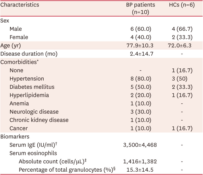
Methods: A case-control study was conducted on 10 newly diagnosed, treatment-naive BP patients and six healthy controls. Disease activity was assessed using the Bullous Pemphigoid Disease Area Index (BPDAI). Peripheral blood mononuclear cells were isolated for FACS analysis to determine immune cell subsets. RNA sequencing was performed to identify differentially expressed genes (DEGs) and enriched pathways. Statistical analyses included t-tests, Mann-Whitney U tests, and correlation analysis.
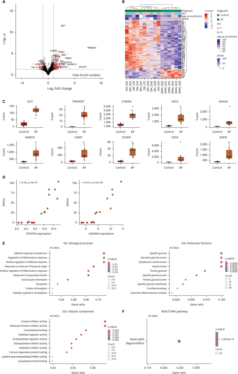
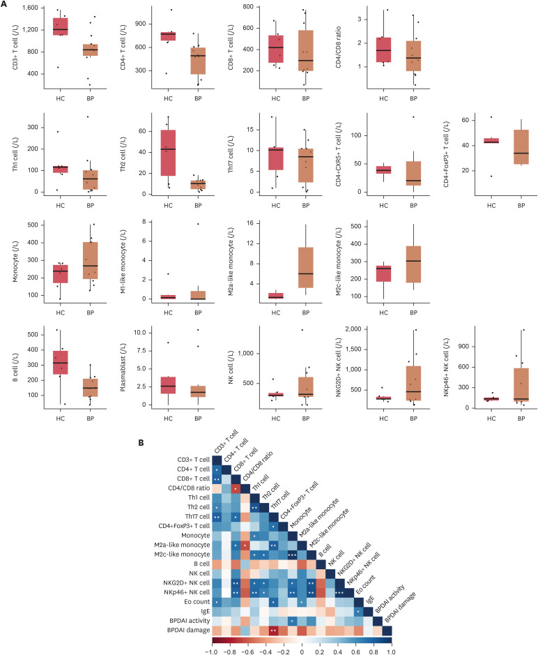
Results: FACS analysis revealed a reduction in CD4⁺ T cells, T helper 2 (Th2), and B cells in BP patients (p<0.05), alongside an increase in M2a-like monocytes (p<0.001). RNA sequencing identified 262 DEGs, with secretory leukocyte peptidase inhibitor (SLPI) and transmembrane protein 237 being the most significantly upregulated. Proline-serine-threonine phosphatase interacting protein 2 (PSTPIP2) and SAM domain, SH3 domain, and nuclear localization signals 1 (SAMSN1) positively correlated with BPDAI (p<0.001). Gene ontology analysis highlighted enrichment in inflammatory responses and neutrophil degranulation pathways.
Conclusion: BP patients show distinct immune dysregulation, including decreased CD4⁺ T cells, Th2, and B cells, increased M2a-like monocytes, altered gene expression profiles, and correlations between PSTPIP2, SAMSN1 and disease activity. These findings provide insights into pathogenesis and potential therapeutic targets of BP.

 求助内容:
求助内容: 应助结果提醒方式:


