{"title":"Metabolic Imaging Parameters of <sup>18</sup>F FDG PET/CT in Differentiating the Ki-67 Index in Carcinoma Breast.","authors":"Deepanksha Datta, Alok Mandal, Akhil Dhanesh Goel, Rajesh Kumar","doi":"10.4103/ijnm.ijnm_168_24","DOIUrl":null,"url":null,"abstract":"<p><strong>Background: </strong>The proliferation index (Ki-67 index) is a known independent prognostic marker in carcinoma breast, and its expression is directly proportional to higher recurrence and worse prognosis. However, there is no standard cutoff of the Ki-67 index to determine its low or high expression. In this study, we aim to find the association of various metabolic parameters on <sup>18</sup>F FDG positron emission tomography/computed tomography (PET/CT) with Ki-67 index in carcinoma breast patients and further evaluate its correlation with low and high Ki-67 index groups.</p><p><strong>Materials and methods: </strong>This is a retrospective study conducted in a tertiary hospital in North India between February 2021 and 2024. All histopathologically proven female cases of carcinoma breast with reported Ki-67 index and baseline <sup>18</sup>F FDG PET/CT before any treatment or surgery were included. The metabolic parameters, namely, standardized uptake value (SUVmax), the metabolic ratio of the primary tumor to the liver (SUR), total lesion glycolysis (TLG), and metabolic tumor volume (MTV) were recorded for each case. The correlation between the metabolic parameters and the Ki-67 index was analyzed, and subgroup analysis was done.</p><p><strong>Results: </strong>Sixty-five female patients met the inclusion criteria, and the majority of them presented with intraductal carcinoma. The median value (interquartile range [IQR]) of the Ki-67 index was 40% (IQR: 50%). The primary breast tumor showed median (IQR) of SUVmax, SUR, TLG, and MTV of 10.3 g/mL (7.2), 3.7 (2.9), 102.9 g.cub/mL (184.7), and 16.2 cm<sup>3</sup> (25.4), respectively. A significant correlation was noted between all the metabolic parameters studied and the Ki-67 index. In subgroup analysis, a significant difference was noted in all the metabolic parameters between the subgroups of the Ki-67 index ≤25% versus >25%.</p><p><strong>Conclusion: </strong>Metabolic parameters on <sup>18</sup>F FDG PET/CT show a promising role in the determination of the status of proliferation marker Ki-67 index in carcinoma breast.</p>","PeriodicalId":45830,"journal":{"name":"Indian Journal of Nuclear Medicine","volume":"40 2","pages":"67-71"},"PeriodicalIF":0.5000,"publicationDate":"2025-03-01","publicationTypes":"Journal Article","fieldsOfStudy":null,"isOpenAccess":false,"openAccessPdf":"https://www.ncbi.nlm.nih.gov/pmc/articles/PMC12303211/pdf/","citationCount":"0","resultStr":null,"platform":"Semanticscholar","paperid":null,"PeriodicalName":"Indian Journal of Nuclear Medicine","FirstCategoryId":"1085","ListUrlMain":"https://doi.org/10.4103/ijnm.ijnm_168_24","RegionNum":0,"RegionCategory":null,"ArticlePicture":[],"TitleCN":null,"AbstractTextCN":null,"PMCID":null,"EPubDate":"2025/6/27 0:00:00","PubModel":"Epub","JCR":"Q4","JCRName":"RADIOLOGY, NUCLEAR MEDICINE & MEDICAL IMAGING","Score":null,"Total":0}
引用次数: 0
Abstract
Background: The proliferation index (Ki-67 index) is a known independent prognostic marker in carcinoma breast, and its expression is directly proportional to higher recurrence and worse prognosis. However, there is no standard cutoff of the Ki-67 index to determine its low or high expression. In this study, we aim to find the association of various metabolic parameters on 18F FDG positron emission tomography/computed tomography (PET/CT) with Ki-67 index in carcinoma breast patients and further evaluate its correlation with low and high Ki-67 index groups.
Materials and methods: This is a retrospective study conducted in a tertiary hospital in North India between February 2021 and 2024. All histopathologically proven female cases of carcinoma breast with reported Ki-67 index and baseline 18F FDG PET/CT before any treatment or surgery were included. The metabolic parameters, namely, standardized uptake value (SUVmax), the metabolic ratio of the primary tumor to the liver (SUR), total lesion glycolysis (TLG), and metabolic tumor volume (MTV) were recorded for each case. The correlation between the metabolic parameters and the Ki-67 index was analyzed, and subgroup analysis was done.
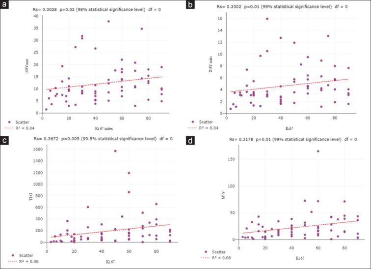
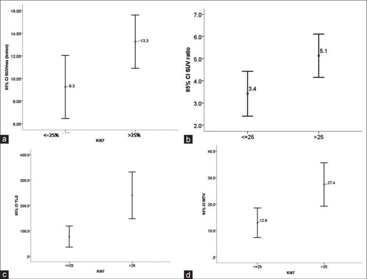
Results: Sixty-five female patients met the inclusion criteria, and the majority of them presented with intraductal carcinoma. The median value (interquartile range [IQR]) of the Ki-67 index was 40% (IQR: 50%). The primary breast tumor showed median (IQR) of SUVmax, SUR, TLG, and MTV of 10.3 g/mL (7.2), 3.7 (2.9), 102.9 g.cub/mL (184.7), and 16.2 cm3 (25.4), respectively. A significant correlation was noted between all the metabolic parameters studied and the Ki-67 index. In subgroup analysis, a significant difference was noted in all the metabolic parameters between the subgroups of the Ki-67 index ≤25% versus >25%.
Conclusion: Metabolic parameters on 18F FDG PET/CT show a promising role in the determination of the status of proliferation marker Ki-67 index in carcinoma breast.

 求助内容:
求助内容: 应助结果提醒方式:


