Effect of Deep Learning-Based Artificial Intelligence on Radiologists' Performance in Identifying Nigrosome 1 Abnormalities on Susceptibility Map-Weighted Imaging.
IF 5.3 2区 医学Q1 RADIOLOGY, NUCLEAR MEDICINE & MEDICAL IMAGING
Jiyeon Park, Chae Young Lim, So Yeon Won, Han Kyu Na, Phil Hyu Lee, Sun-Young Baek, Yun Hwa Roh, Minjung Seong, Yongsik Sim, Eung Yeop Kim, Sung Tae Kim, Beomseok Sohn
{"title":"Effect of Deep Learning-Based Artificial Intelligence on Radiologists' Performance in Identifying Nigrosome 1 Abnormalities on Susceptibility Map-Weighted Imaging.","authors":"Jiyeon Park, Chae Young Lim, So Yeon Won, Han Kyu Na, Phil Hyu Lee, Sun-Young Baek, Yun Hwa Roh, Minjung Seong, Yongsik Sim, Eung Yeop Kim, Sung Tae Kim, Beomseok Sohn","doi":"10.3348/kjr.2025.0208","DOIUrl":null,"url":null,"abstract":"<p><strong>Objective: </strong>To evaluate the effect of deep learning (DL)-based artificial intelligence (AI) software on the diagnostic performance of radiologists with different experience levels in detecting nigrosome 1 (N1) abnormalities on susceptibility map-weighted imaging (SMwI).</p><p><strong>Materials and methods: </strong>This retrospective diagnostic case-control study analyzed 139 SMwI scans of 59 patients with Parkinson's disease (PD) and 80 healthy participants. Participants were imaged using 3T MRI, and AI-generated assessments for N1 abnormalities were obtained using an AI model (version 1.0.1.0; Heuron Corporation, Seoul, Korea), which utilized YOLOX-based object detection and SparseInst segmentation models. Four radiologists (two experienced neuroradiologists and two less experienced residents) evaluated N1 abnormalities with and without AI in a crossover study design. Diagnostic performance metrics, inter-reader agreements, and reader responses to AI-generated assessments were evaluated.</p><p><strong>Results: </strong>Use of AI significantly improved diagnostic performance compared with interpretation without it across three readers, with significant increases in specificity (0.86 vs. 0.94, <i>P</i> = 0.004; 0.91 vs. 0.97, <i>P</i> = 0.024; and 0.90 vs. 0.97, <i>P</i> = 0.012). Inter-reader agreement also improved with AI, as Fleiss's kappa increased from 0.73 (95% confidence interval [CI]: 0.61-0.84) to 0.87 (95% CI: 0.76-0.99). The net reclassification index (NRI) demonstrated significant improvement in three of the four readers. When grouped by experience level, less experienced readers showed greater improvement (NRI = 12.8%, 95% CI: 0.067-0.190) than experienced readers (NRI = 0.8%, 95% CI: -0.037-0.051). In the less experienced group, reader-AI disagreement was significantly higher in the PD group than in the normal group (8.1% vs. 3.8%, <i>P</i> = 0.029).</p><p><strong>Conclusion: </strong>DL-based AI enhances the diagnostic performance in detecting N1 abnormalities on SMwI, particularly benefiting less experienced radiologists. These findings underscore the potential for improving diagnostic workflows for PD.</p>","PeriodicalId":17881,"journal":{"name":"Korean Journal of Radiology","volume":"26 8","pages":"771-781"},"PeriodicalIF":5.3000,"publicationDate":"2025-08-01","publicationTypes":"Journal Article","fieldsOfStudy":null,"isOpenAccess":false,"openAccessPdf":"https://www.ncbi.nlm.nih.gov/pmc/articles/PMC12318656/pdf/","citationCount":"0","resultStr":null,"platform":"Semanticscholar","paperid":null,"PeriodicalName":"Korean Journal of Radiology","FirstCategoryId":"3","ListUrlMain":"https://doi.org/10.3348/kjr.2025.0208","RegionNum":2,"RegionCategory":"医学","ArticlePicture":[],"TitleCN":null,"AbstractTextCN":null,"PMCID":null,"EPubDate":"","PubModel":"","JCR":"Q1","JCRName":"RADIOLOGY, NUCLEAR MEDICINE & MEDICAL IMAGING","Score":null,"Total":0}
引用次数: 0
Abstract
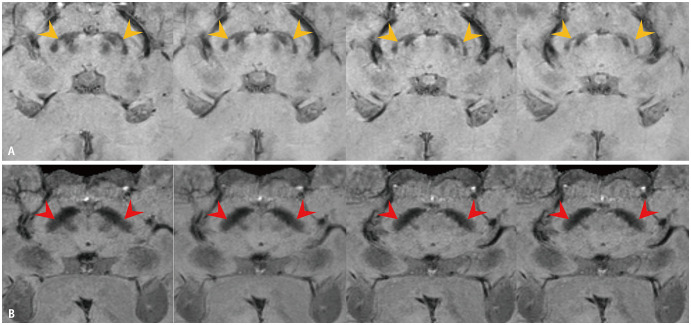
Objective: To evaluate the effect of deep learning (DL)-based artificial intelligence (AI) software on the diagnostic performance of radiologists with different experience levels in detecting nigrosome 1 (N1) abnormalities on susceptibility map-weighted imaging (SMwI).
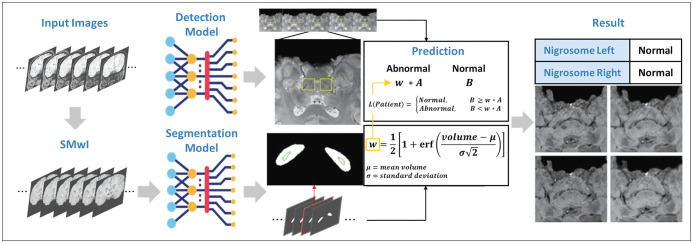
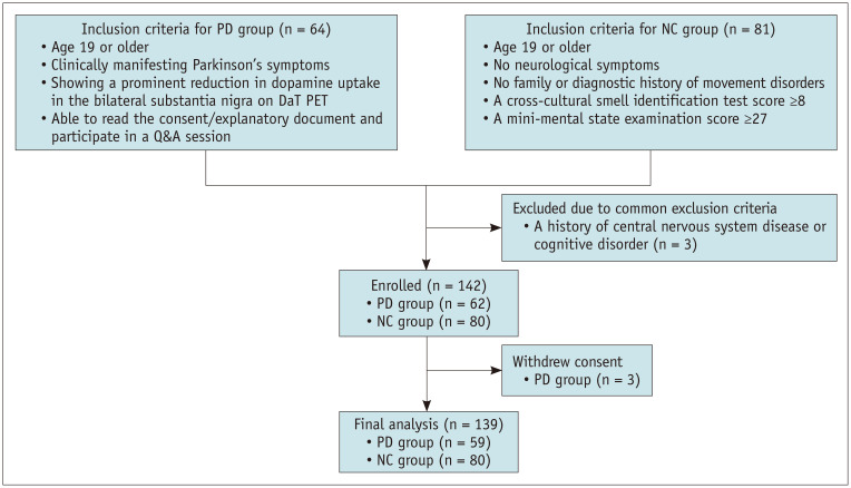
Materials and methods: This retrospective diagnostic case-control study analyzed 139 SMwI scans of 59 patients with Parkinson's disease (PD) and 80 healthy participants. Participants were imaged using 3T MRI, and AI-generated assessments for N1 abnormalities were obtained using an AI model (version 1.0.1.0; Heuron Corporation, Seoul, Korea), which utilized YOLOX-based object detection and SparseInst segmentation models. Four radiologists (two experienced neuroradiologists and two less experienced residents) evaluated N1 abnormalities with and without AI in a crossover study design. Diagnostic performance metrics, inter-reader agreements, and reader responses to AI-generated assessments were evaluated.
Results: Use of AI significantly improved diagnostic performance compared with interpretation without it across three readers, with significant increases in specificity (0.86 vs. 0.94, P = 0.004; 0.91 vs. 0.97, P = 0.024; and 0.90 vs. 0.97, P = 0.012). Inter-reader agreement also improved with AI, as Fleiss's kappa increased from 0.73 (95% confidence interval [CI]: 0.61-0.84) to 0.87 (95% CI: 0.76-0.99). The net reclassification index (NRI) demonstrated significant improvement in three of the four readers. When grouped by experience level, less experienced readers showed greater improvement (NRI = 12.8%, 95% CI: 0.067-0.190) than experienced readers (NRI = 0.8%, 95% CI: -0.037-0.051). In the less experienced group, reader-AI disagreement was significantly higher in the PD group than in the normal group (8.1% vs. 3.8%, P = 0.029).
Conclusion: DL-based AI enhances the diagnostic performance in detecting N1 abnormalities on SMwI, particularly benefiting less experienced radiologists. These findings underscore the potential for improving diagnostic workflows for PD.
期刊介绍:
The inaugural issue of the Korean J Radiol came out in March 2000. Our journal aims to produce and propagate knowledge on radiologic imaging and related sciences.
A unique feature of the articles published in the Journal will be their reflection of global trends in radiology combined with an East-Asian perspective. Geographic differences in disease prevalence will be reflected in the contents of papers, and this will serve to enrich our body of knowledge.
World''s outstanding radiologists from many countries are serving as editorial board of our journal.

 求助内容:
求助内容: 应助结果提醒方式:


