Challenges and predictive value of morphologic features in panfungal sequencing of formalin-fixed, paraffin-embedded tissues: A 5-year retrospective study.
Shane A Kaysen, Matt X Luo, Ryan C Shean, Kimberly E Hanson, Benjamin T Bradley, Gillian L Hale
{"title":"Challenges and predictive value of morphologic features in panfungal sequencing of formalin-fixed, paraffin-embedded tissues: A 5-year retrospective study.","authors":"Shane A Kaysen, Matt X Luo, Ryan C Shean, Kimberly E Hanson, Benjamin T Bradley, Gillian L Hale","doi":"10.1093/ajcp/aqaf073","DOIUrl":null,"url":null,"abstract":"<p><strong>Objective: </strong>Panfungal sequencing (PFS) using formalin-fixed, paraffin-embedded (FFPE) tissue aids genus-level or species-level identification in suspected invasive fungal infections. Given the limited availability of PFS and potential risk of environmental contamination, defining histopathologic features predictive of clinically interpretable results is important.</p><p><strong>Methods: </strong>We evaluated FFPE tissue samples submitted for PFS over a 5-year period. Histopathologic data were extracted from pathology reports; in-house cases were re-reviewed, and the burden of fungal elements was assessed using Grocott methenamine silver stain. Any available fungal culture data were also obtained for in-house cases.</p><p><strong>Results: </strong>Of 153 cases with fungal elements reported by histopathology, 54% were positive by PFS. Of 97 cases without histologic evidence of fungal elements, only 9% were positive by PFS, and all were considered potential environmental contaminants. Culture results were available for only 9 of 461 (2%) cases, and all cultures were concordant with the PFS results. When the pathologist proposed 1 or more specific organisms based on histologic appearance alone, PFS was discrepant in 37% of cases. Of those discrepant diagnoses, and if we designate the PFS result as the true diagnosis, then 53% of misclassifications had the potential for administration of suboptimal antifungal therapy. There was no correlation between the relative abundance of fungal elements in tissue sections and detection of fungal organisms by PFS.</p><p><strong>Conclusions: </strong>Panfungal sequencing can provide genus-level and species-level identification in the setting of visible fungal elements in FFPE tissue. It is a valuable diagnostic tool, particularly when fungal infections are clinically suspected but fungal cultures were not performed.</p>","PeriodicalId":7506,"journal":{"name":"American journal of clinical pathology","volume":" ","pages":"464-473"},"PeriodicalIF":1.9000,"publicationDate":"2025-09-09","publicationTypes":"Journal Article","fieldsOfStudy":null,"isOpenAccess":false,"openAccessPdf":"https://www.ncbi.nlm.nih.gov/pmc/articles/PMC12421234/pdf/","citationCount":"0","resultStr":null,"platform":"Semanticscholar","paperid":null,"PeriodicalName":"American journal of clinical pathology","FirstCategoryId":"3","ListUrlMain":"https://doi.org/10.1093/ajcp/aqaf073","RegionNum":4,"RegionCategory":"医学","ArticlePicture":[],"TitleCN":null,"AbstractTextCN":null,"PMCID":null,"EPubDate":"","PubModel":"","JCR":"Q2","JCRName":"PATHOLOGY","Score":null,"Total":0}
引用次数: 0
Abstract
Objective: Panfungal sequencing (PFS) using formalin-fixed, paraffin-embedded (FFPE) tissue aids genus-level or species-level identification in suspected invasive fungal infections. Given the limited availability of PFS and potential risk of environmental contamination, defining histopathologic features predictive of clinically interpretable results is important.
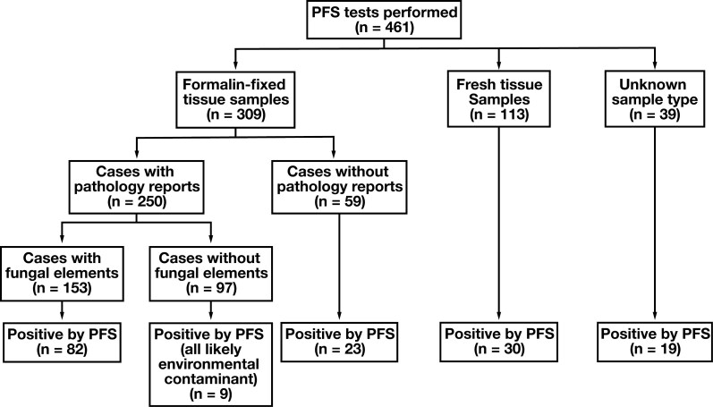
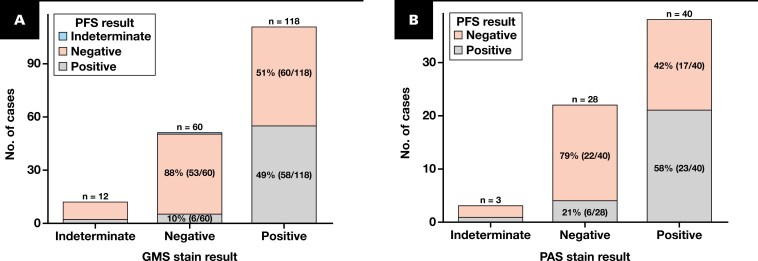
Methods: We evaluated FFPE tissue samples submitted for PFS over a 5-year period. Histopathologic data were extracted from pathology reports; in-house cases were re-reviewed, and the burden of fungal elements was assessed using Grocott methenamine silver stain. Any available fungal culture data were also obtained for in-house cases.
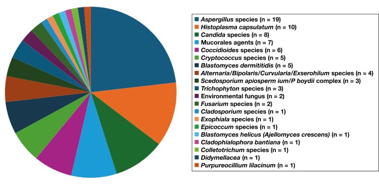
Results: Of 153 cases with fungal elements reported by histopathology, 54% were positive by PFS. Of 97 cases without histologic evidence of fungal elements, only 9% were positive by PFS, and all were considered potential environmental contaminants. Culture results were available for only 9 of 461 (2%) cases, and all cultures were concordant with the PFS results. When the pathologist proposed 1 or more specific organisms based on histologic appearance alone, PFS was discrepant in 37% of cases. Of those discrepant diagnoses, and if we designate the PFS result as the true diagnosis, then 53% of misclassifications had the potential for administration of suboptimal antifungal therapy. There was no correlation between the relative abundance of fungal elements in tissue sections and detection of fungal organisms by PFS.
Conclusions: Panfungal sequencing can provide genus-level and species-level identification in the setting of visible fungal elements in FFPE tissue. It is a valuable diagnostic tool, particularly when fungal infections are clinically suspected but fungal cultures were not performed.
期刊介绍:
The American Journal of Clinical Pathology (AJCP) is the official journal of the American Society for Clinical Pathology and the Academy of Clinical Laboratory Physicians and Scientists. It is a leading international journal for publication of articles concerning novel anatomic pathology and laboratory medicine observations on human disease. AJCP emphasizes articles that focus on the application of evolving technologies for the diagnosis and characterization of diseases and conditions, as well as those that have a direct link toward improving patient care.
 求助内容:
求助内容: 应助结果提醒方式:


