Adib Al-Haj Husain, Peter Kessler, Suen An Nynke Lie, Samuel Drack, Egon Burian, Sameena Sandhu, Maximilian Eberhard Hermann Wagner, Bernd Stadlinger, Thomas Frauenfelder, Giovanni Colacicco, Rubens Spin-Neto, Harald Essig
{"title":"Black bone and CT-like MRI-based delineation of fracture-prone regions in oral and maxillofacial trauma.","authors":"Adib Al-Haj Husain, Peter Kessler, Suen An Nynke Lie, Samuel Drack, Egon Burian, Sameena Sandhu, Maximilian Eberhard Hermann Wagner, Bernd Stadlinger, Thomas Frauenfelder, Giovanni Colacicco, Rubens Spin-Neto, Harald Essig","doi":"10.1007/s10006-025-01431-6","DOIUrl":null,"url":null,"abstract":"<p><strong>Purpose: </strong>To assess the effectiveness and feasibility of MRI-based delineation of fracture-prone regions in the oral and maxillofacial region using Black Bone and CT-like MRI protocols optimized for dentomaxillofacial imaging with a dedicated 15-channel mandibular coil.</p><p><strong>Methods: </strong>In this prospective study, healthy volunteers underwent 3T MRI using five protocols: DESS, SPACE STIR, SPACE SPAIR, T1-VIBE Dixon, and UTE. Eight trauma-prone regions, including the nasal septum, orbit, naso-orbito-ethmoidal (NOE) complex, zygomaticomaxillary complex, Le Fort regions, mandible, condyle, and dentoalveolar complex, were assessed. Image quality, artifacts, anatomical delineation, and bone-to-soft-tissue contrast were rated on a five-point Likert scale by three independent observers. Descriptive statistics and inter-rater agreement (intraclass correlation coefficients (ICCs)) were calculated.</p><p><strong>Results: </strong>Sixteen participants (37.2 ± 12.9 years; 12 males, 4 females) were included, resulting in 80 MRI volumes and 640 regions for evaluation. UTE and VIBE-DIXON sequences achieved the highest ratings for image quality, artifact susceptibility, and anatomical delineation across most fracture-prone regions (ICC: 0.793-1; all p < 0.001). UTE excelled in visualizing NOE and Le Fort regions, while VIBE-DIXON performed best in mandibular and orbital imaging. Bone-to-soft-tissue contrast was highest in UTE and VIBE-DIXON, highlighting their diagnostic potential in simultaneous soft and hard tissue imaging. Inter-rater agreement was consistently high (ICC: 0.772-0.976; all p < 0.001).</p><p><strong>Conclusion: </strong>Dedicated trauma-specific MRI protocols show promising potential as a radiation-free modality for maxillofacial trauma imaging, particularly in young adults and pediatric patients. The strengths of each protocol highlight the need for tailored sequence selection to optimize diagnostic accuracy and personalized care.</p><p><strong>Trial registration number: </strong>Swiss National Clinical Trials Portal: SNCTP000005246.</p>","PeriodicalId":520733,"journal":{"name":"Oral and maxillofacial surgery","volume":"29 1","pages":"136"},"PeriodicalIF":1.8000,"publicationDate":"2025-07-28","publicationTypes":"Journal Article","fieldsOfStudy":null,"isOpenAccess":false,"openAccessPdf":"https://www.ncbi.nlm.nih.gov/pmc/articles/PMC12304013/pdf/","citationCount":"0","resultStr":null,"platform":"Semanticscholar","paperid":null,"PeriodicalName":"Oral and maxillofacial surgery","FirstCategoryId":"1085","ListUrlMain":"https://doi.org/10.1007/s10006-025-01431-6","RegionNum":0,"RegionCategory":null,"ArticlePicture":[],"TitleCN":null,"AbstractTextCN":null,"PMCID":null,"EPubDate":"","PubModel":"","JCR":"","JCRName":"","Score":null,"Total":0}
引用次数: 0
Abstract
Purpose: To assess the effectiveness and feasibility of MRI-based delineation of fracture-prone regions in the oral and maxillofacial region using Black Bone and CT-like MRI protocols optimized for dentomaxillofacial imaging with a dedicated 15-channel mandibular coil.
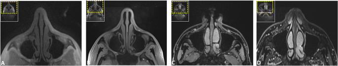
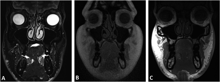
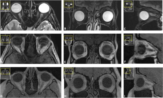
Methods: In this prospective study, healthy volunteers underwent 3T MRI using five protocols: DESS, SPACE STIR, SPACE SPAIR, T1-VIBE Dixon, and UTE. Eight trauma-prone regions, including the nasal septum, orbit, naso-orbito-ethmoidal (NOE) complex, zygomaticomaxillary complex, Le Fort regions, mandible, condyle, and dentoalveolar complex, were assessed. Image quality, artifacts, anatomical delineation, and bone-to-soft-tissue contrast were rated on a five-point Likert scale by three independent observers. Descriptive statistics and inter-rater agreement (intraclass correlation coefficients (ICCs)) were calculated.
Results: Sixteen participants (37.2 ± 12.9 years; 12 males, 4 females) were included, resulting in 80 MRI volumes and 640 regions for evaluation. UTE and VIBE-DIXON sequences achieved the highest ratings for image quality, artifact susceptibility, and anatomical delineation across most fracture-prone regions (ICC: 0.793-1; all p < 0.001). UTE excelled in visualizing NOE and Le Fort regions, while VIBE-DIXON performed best in mandibular and orbital imaging. Bone-to-soft-tissue contrast was highest in UTE and VIBE-DIXON, highlighting their diagnostic potential in simultaneous soft and hard tissue imaging. Inter-rater agreement was consistently high (ICC: 0.772-0.976; all p < 0.001).
Conclusion: Dedicated trauma-specific MRI protocols show promising potential as a radiation-free modality for maxillofacial trauma imaging, particularly in young adults and pediatric patients. The strengths of each protocol highlight the need for tailored sequence selection to optimize diagnostic accuracy and personalized care.
Trial registration number: Swiss National Clinical Trials Portal: SNCTP000005246.

 求助内容:
求助内容: 应助结果提醒方式:


