{"title":"Uncommon presentation of dermatofibrosarcoma protuberans: extensive growth in a rare location with subclinical cutaneous manifestation-a case report.","authors":"Zain Elabedin Asheer, Jennifer Berg Drejøe","doi":"10.1080/23320885.2025.2535699","DOIUrl":null,"url":null,"abstract":"<p><p>Dermatofibrosarcoma protuberans (DFSP) is a rare, locally aggressive cutaneous tumor predominantly affecting young to middle-aged adults, characterized by a slow-growing, indurated plaque or nodule. The diagnosis and treatment of DFSP can be challenging due to its rarity, growth pattern and variable clinical presentation. A 40-year-old male presented with a firm lump of 8 mm on his forehead for one year. It was initially suspected to be a benign lipoma or atheroma and removed accordingly by a private plastic surgeon. However, histopathological examination revealed dermatofibrosarcoma protuberans (DFSP) that was not radically removed, prompting referral for removal at our department. Here, he presented with a scar of 10 mm with no visible or palpable residual tumor. A planned excision of 20 mm and reconstruction with split thickness skin graft (STSG) was performed. Histopathology showed tumor activity at several margins, with perineural and periosteal invasion. MRI was inconclusive, and therefore PET-CT was added, which showed possible residual tumor. Re-excision including mapping biopsies was performed. Wide re-excision was performed three times to achieve clear surgical margins, yielding a tumor size of approximately 8x10 cm and a final defect measuring 10x12 cm. The defect was reconstructed with a dermal template (Integra<sup>®</sup>) and STSG. Our case underscores the propensity of DFSP for significant subclinical extension, including potential perineural and periosteal invasion. Despite the aesthetically challenging location, a satisfactory cosmetic result was achieved, and there was no recurrence during the two-year follow-up.</p>","PeriodicalId":42421,"journal":{"name":"Case Reports in Plastic Surgery and Hand Surgery","volume":"12 1","pages":"2535699"},"PeriodicalIF":0.6000,"publicationDate":"2025-07-26","publicationTypes":"Journal Article","fieldsOfStudy":null,"isOpenAccess":false,"openAccessPdf":"https://www.ncbi.nlm.nih.gov/pmc/articles/PMC12302385/pdf/","citationCount":"0","resultStr":null,"platform":"Semanticscholar","paperid":null,"PeriodicalName":"Case Reports in Plastic Surgery and Hand Surgery","FirstCategoryId":"1085","ListUrlMain":"https://doi.org/10.1080/23320885.2025.2535699","RegionNum":0,"RegionCategory":null,"ArticlePicture":[],"TitleCN":null,"AbstractTextCN":null,"PMCID":null,"EPubDate":"2025/1/1 0:00:00","PubModel":"eCollection","JCR":"Q4","JCRName":"SURGERY","Score":null,"Total":0}
引用次数: 0
Abstract
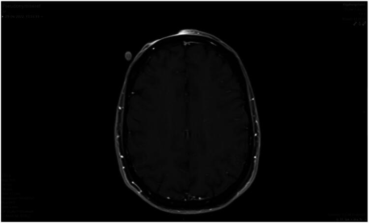
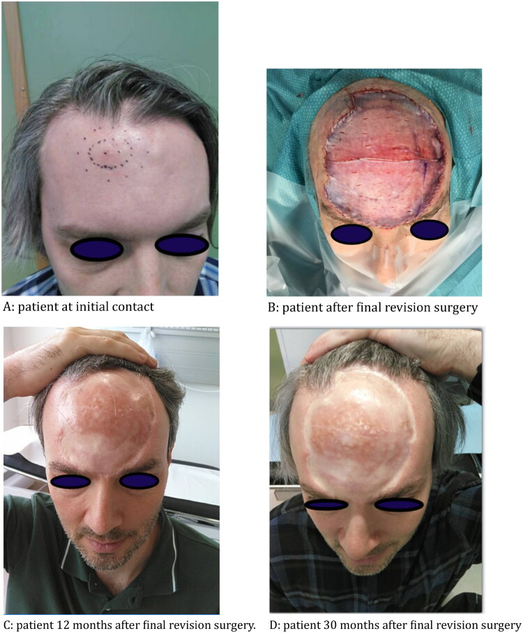
Dermatofibrosarcoma protuberans (DFSP) is a rare, locally aggressive cutaneous tumor predominantly affecting young to middle-aged adults, characterized by a slow-growing, indurated plaque or nodule. The diagnosis and treatment of DFSP can be challenging due to its rarity, growth pattern and variable clinical presentation. A 40-year-old male presented with a firm lump of 8 mm on his forehead for one year. It was initially suspected to be a benign lipoma or atheroma and removed accordingly by a private plastic surgeon. However, histopathological examination revealed dermatofibrosarcoma protuberans (DFSP) that was not radically removed, prompting referral for removal at our department. Here, he presented with a scar of 10 mm with no visible or palpable residual tumor. A planned excision of 20 mm and reconstruction with split thickness skin graft (STSG) was performed. Histopathology showed tumor activity at several margins, with perineural and periosteal invasion. MRI was inconclusive, and therefore PET-CT was added, which showed possible residual tumor. Re-excision including mapping biopsies was performed. Wide re-excision was performed three times to achieve clear surgical margins, yielding a tumor size of approximately 8x10 cm and a final defect measuring 10x12 cm. The defect was reconstructed with a dermal template (Integra®) and STSG. Our case underscores the propensity of DFSP for significant subclinical extension, including potential perineural and periosteal invasion. Despite the aesthetically challenging location, a satisfactory cosmetic result was achieved, and there was no recurrence during the two-year follow-up.

 求助内容:
求助内容: 应助结果提醒方式:


