{"title":"Lung ultrasound findings and therapeutic outcome in a cat with recurrent spontaneous pneumothorax caused by paragonimiasis.","authors":"Po-Yao Huang, Chi-Ru Chen, Chung-Hui Lin, Pei-Ying Lo, Ju-Hsien Peng, Fei-Hsuan Wang, Olivia F Hsieh, Hui-Wen Chen, Wei-Tao Chang","doi":"10.1177/20551169251341545","DOIUrl":null,"url":null,"abstract":"<p><strong>Case summary: </strong>A 1-year-old intact female domestic shorthair cat presented for evaluation of worsening respiratory distress and tachypnoea. The cat had been diagnosed with spontaneous pneumothorax 4 days earlier by the primary clinician and treated with therapeutic thoracocentesis. On physical examination, the patient exhibited decreased lung sounds, tachypnoea and increased breathing effort. Recurrent spontaneous pneumothorax was confirmed via thoracic radiography, and therapeutic thoracocentesis was repeated. Lung ultrasound performed after thoracocentesis revealed lung consolidation, pulmonary nodules and multiple cyst-like lesions with irregularly thickened echogenic walls and anechoic centres. Repeat thoracic radiography showed a diffuse moderate bronchointerstitial pattern with multifocal soft tissue nodules. A thorough faecal examination revealed ova resembling those of <i>Paragonimus</i> species, and subsequent molecular analysis confirmed <i>Paragonimus westermani</i>. The cat was initially treated with fenbendazole alone, which resulted in limited improvement and recurrent spontaneous pneumothorax. A second course of fenbendazole combined with praziquantel led to clinical improvement. The cat remained free of clinical signs and was doing well, with no identifiable lung nodules on thoracic radiography 2 years after diagnosis.</p><p><strong>Relevance and novel information: </strong>This report describes a novel cyst-like lung ultrasound finding characterised by an irregularly thickened echogenic wall and anechoic centre, associated with <i>P westermani</i> infection in a cat presenting with recurrent spontaneous pneumothorax. The report also highlights a successful treatment approach resulting in long-term resolution. The identification of this novel lung ultrasound finding can facilitate early diagnosis and treatment of <i>Paragonimus</i> species infection in cats, especially for those presenting with respiratory distress and pneumothorax.</p>","PeriodicalId":36588,"journal":{"name":"Journal of Feline Medicine and Surgery Open Reports","volume":"11 2","pages":"20551169251341545"},"PeriodicalIF":0.7000,"publicationDate":"2025-07-22","publicationTypes":"Journal Article","fieldsOfStudy":null,"isOpenAccess":false,"openAccessPdf":"https://www.ncbi.nlm.nih.gov/pmc/articles/PMC12290250/pdf/","citationCount":"0","resultStr":null,"platform":"Semanticscholar","paperid":null,"PeriodicalName":"Journal of Feline Medicine and Surgery Open Reports","FirstCategoryId":"1085","ListUrlMain":"https://doi.org/10.1177/20551169251341545","RegionNum":0,"RegionCategory":null,"ArticlePicture":[],"TitleCN":null,"AbstractTextCN":null,"PMCID":null,"EPubDate":"2025/7/1 0:00:00","PubModel":"eCollection","JCR":"Q3","JCRName":"VETERINARY SCIENCES","Score":null,"Total":0}
引用次数: 0
Abstract
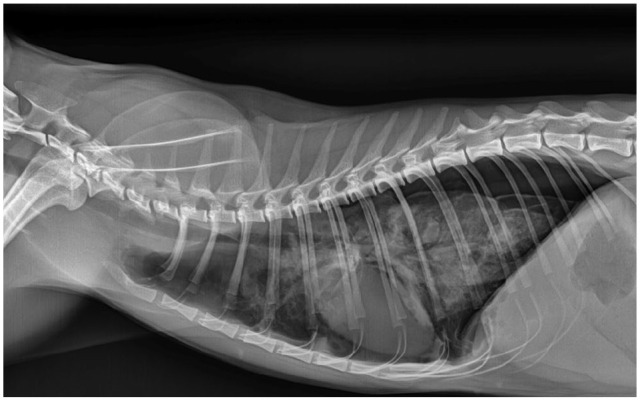
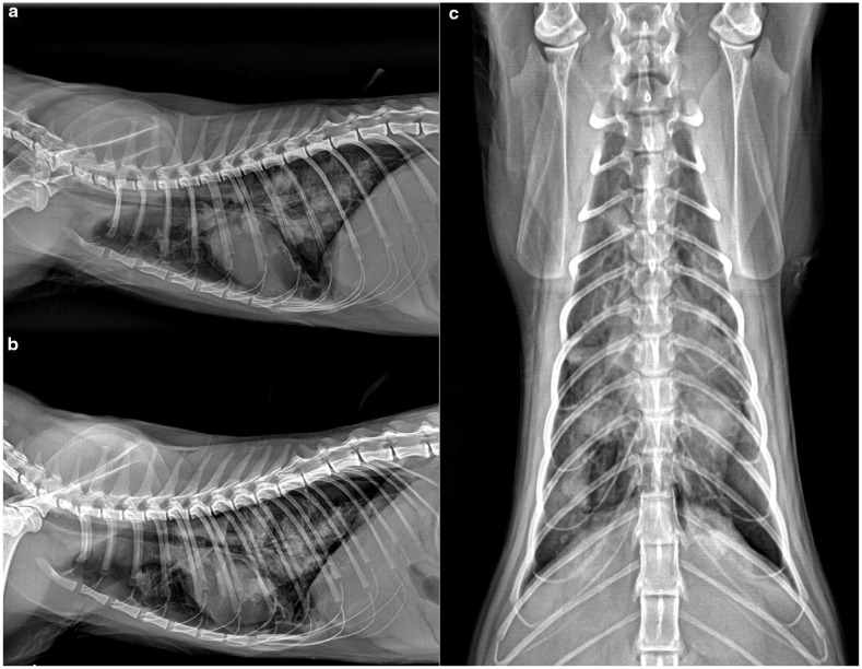
Case summary: A 1-year-old intact female domestic shorthair cat presented for evaluation of worsening respiratory distress and tachypnoea. The cat had been diagnosed with spontaneous pneumothorax 4 days earlier by the primary clinician and treated with therapeutic thoracocentesis. On physical examination, the patient exhibited decreased lung sounds, tachypnoea and increased breathing effort. Recurrent spontaneous pneumothorax was confirmed via thoracic radiography, and therapeutic thoracocentesis was repeated. Lung ultrasound performed after thoracocentesis revealed lung consolidation, pulmonary nodules and multiple cyst-like lesions with irregularly thickened echogenic walls and anechoic centres. Repeat thoracic radiography showed a diffuse moderate bronchointerstitial pattern with multifocal soft tissue nodules. A thorough faecal examination revealed ova resembling those of Paragonimus species, and subsequent molecular analysis confirmed Paragonimus westermani. The cat was initially treated with fenbendazole alone, which resulted in limited improvement and recurrent spontaneous pneumothorax. A second course of fenbendazole combined with praziquantel led to clinical improvement. The cat remained free of clinical signs and was doing well, with no identifiable lung nodules on thoracic radiography 2 years after diagnosis.
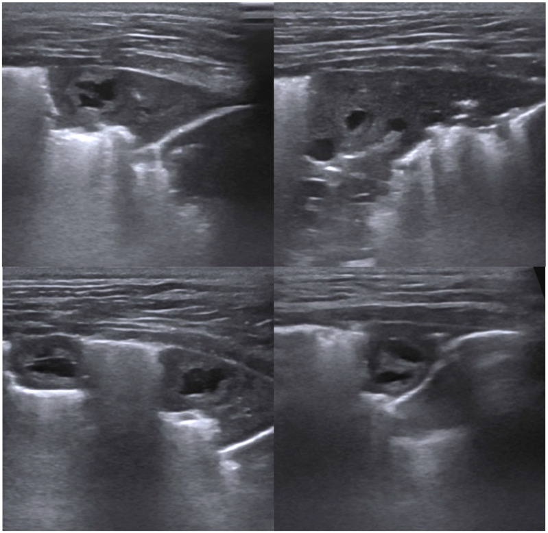
Relevance and novel information: This report describes a novel cyst-like lung ultrasound finding characterised by an irregularly thickened echogenic wall and anechoic centre, associated with P westermani infection in a cat presenting with recurrent spontaneous pneumothorax. The report also highlights a successful treatment approach resulting in long-term resolution. The identification of this novel lung ultrasound finding can facilitate early diagnosis and treatment of Paragonimus species infection in cats, especially for those presenting with respiratory distress and pneumothorax.

 求助内容:
求助内容: 应助结果提醒方式:


